
Mitochondrial Stress Signaling and Disease
Mitochondrial Stress Signaling and Disease
Mitochondrial functional disturbance can activate a series of stress signals to restore energy metabolism, protein homeostasis, and redox balance. Decreased membrane potential, impaired respiratory chain function, defective protein import, or abnormal mitochondrial proteostasis can induce multiple stress responses, including the ISR, UPRmt, mitophagy, and mitochondrial factor secretion. Short-term stress helps cells adapt, whereas long-term or abnormal activation may contribute to aging, tumors, neurodegenerative diseases, metabolic disorders, and chronic inflammation.
Keywords: mitochondrial stress; integrated stress response; DELE1; OMA1; HRI; ATF4; UPRmt; mitochondrial protein import; GDF15; FGF21; mitophagy; mitohormesis; disease mechanisms
1 Research Entry Points for Mitochondrial Stress
1.1 Main Sources of Mitochondrial Stress
Mitochondrial stress is not a single injury event, but an integrated response formed after abnormalities in mitochondrial membrane potential, oxidative phosphorylation, protein import, protein folding, mtDNA stability, and redox status. Different sources of stress can activate different signaling branches, but they often converge on proteostasis, metabolic remodeling, inflammatory regulation, and altered cell fate.
Stress Source | Direct Effect | Main Response Pathway |
Respiratory chain impairment | ATP decline, NADH/NAD⁺ imbalance, ROS elevation | ISR, AMPK, redox response |
Decreased membrane potential | Protein import defects, ion homeostasis disturbance | PINK1/Parkin, OMA1-DELE1-HRI |
Mitochondrial protein misfolding | Impaired matrix proteostasis | UPRmt, mitochondrial protease system |
Blocked mitochondrial protein import | Accumulation of precursor proteins in the cytosol | mPOS, mitoCPR, mitoRQC, etc. |
mtDNA damage or release | Inflammation and immune recognition | cGAS-STING, TLR9, inflammasome |
Increased mitochondrial ROS | Altered redox signaling | NRF2, ISR, mitohormesis |
Abnormal mitochondrial dynamics | Imbalance among fusion, fission, and clearance | DRP1, MFN1/2, OPA1, mitophagy |
1.2 Adaptive and Pathological Responses
Mitochondrial stress has a dual nature. Mild and transient mitochondrial pressure can enhance antioxidant capacity, protein quality control, and metabolic flexibility, representing protective mitohormesis. However, when stress is too strong or persists for too long, the same signals may shift toward pathological effects, promoting inflammation, cell death, tissue functional decline, or tumor adaptation.
Stress Intensity/Duration | Main Result | Biological Significance |
Mild and transient | Metabolic adaptation and enhanced antioxidant capacity | Protective mitohormesis |
Moderate and recoverable | Enhanced ISR, UPRmt, and mitophagy | Homeostatic repair |
Strong and persistent | Proteostasis collapse, inflammation, and cell death | Disease progression |
Chronic low-level | Adaptive cellular reprogramming | Aging, tumor tolerance, metabolic abnormalities |
2 OMA1-DELE1-HRI Axis and the Integrated Stress Response
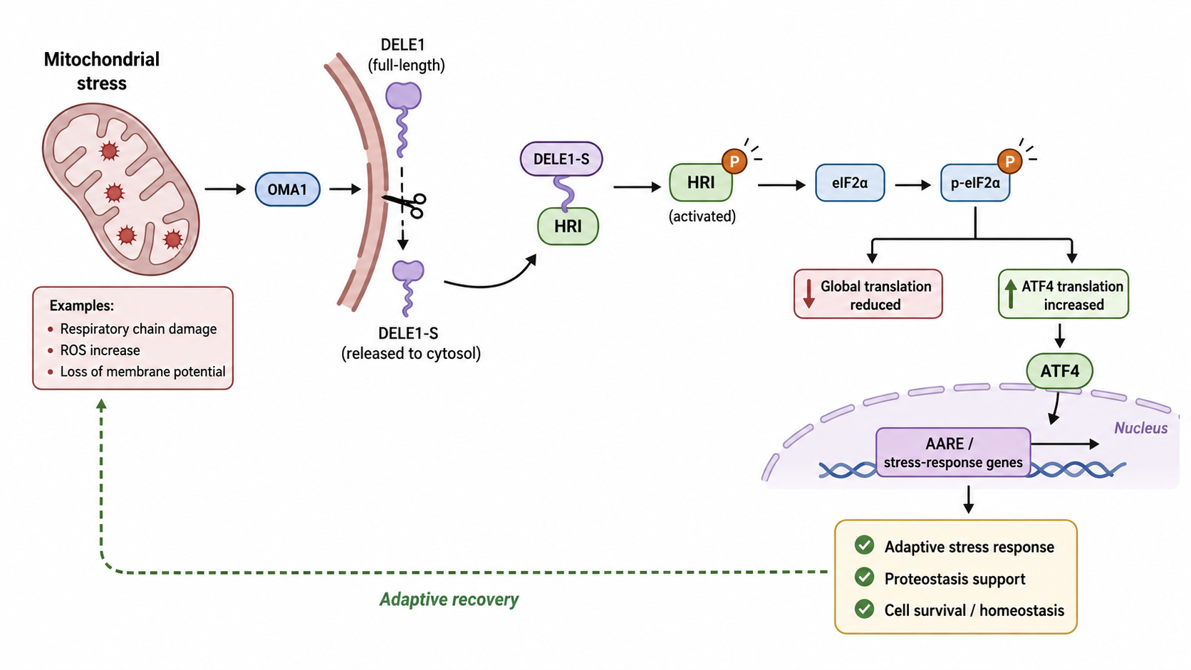
Figure 1. Mitochondrial Integrated Stress Response Mediated by the OMA1-DELE1-HRI-eIF2α-ATF4 Axis
2.1 OMA1 Senses Mitochondrial Functional Disturbance
OMA1 is a stress-sensitive protease located in the mitochondrial inner membrane. When mitochondrial membrane potential decreases, respiratory chain function is impaired, protein import becomes abnormal, or membrane protein homeostasis is disrupted, OMA1 can be activated and cleave mitochondrial-associated substrates. One of its important functions is to mediate DELE1 cleavage, thereby transmitting internal mitochondrial damage signals to the cytosolic ISR pathway.
OMA1 can be understood as a stress-sensing node in the mitochondrial inner membrane. When mitochondria cannot maintain normal membrane potential and protein homeostasis, OMA1 converts local mitochondrial pressure into a transcriptional response that can be sensed by the nucleus.
2.2 DELE1 Connects Mitochondria with Cytosolic Stress Kinases
DELE1 is a key protein in mitochondrial stress transmission. Under normal conditions, DELE1 is localized in mitochondria. When mitochondrial stress occurs, OMA1 can cleave DELE1 to produce a shorter cytosolic form, DELE1-S. After entering the cytosol, DELE1-S can bind and activate HRI.
In addition to OMA1-mediated cleavage, impaired mitochondrial protein import, low iron status, or translocation defects may also cause abnormal accumulation of full-length DELE1 on the outer side of mitochondria or in the cytosol, thereby participating in HRI activation. In other words, DELE1 not only responds to membrane potential-related damage, but also participates in broader stress recognition related to protein import and mitochondrial dysfunction.
2.3 HRI-eIF2α-ATF4 Mediates the ISR Transcriptional Program
After HRI is activated by DELE1, it can phosphorylate eIF2α. eIF2α phosphorylation suppresses global protein translation, but promotes translation of specific transcription factors such as ATF4. ATF4 then induces gene expression related to amino acid metabolism, antioxidant responses, proteostasis regulation, mitochondrial metabolic remodeling, and cell fate.
Node | Function | Result |
Mitochondrial damage | Membrane potential decline, import blockage, proteostasis abnormality | Activates OMA1 or promotes abnormal DELE1 localization |
OMA1 | Cleaves DELE1 | Generates cytosolic DELE1-S |
DELE1 | Activates HRI | Transmits mitochondrial stress to the ISR |
HRI | Phosphorylates eIF2α | Suppresses global translation and alters stress protein synthesis |
ATF4 | Initiates stress transcriptional program | Regulates metabolism, antioxidant responses, proteostasis, and cell fate |
2.4 Protective and Pathological Effects of the ISR
Short-term ISR activation helps reduce newly synthesized protein burden, adjust metabolism, enhance antioxidant capacity, and improve cell survival. If the ISR remains activated for a long period, it may cause excessive translation inhibition, reduced cellular function, increased inflammatory factor expression, or activation of apoptosis/death-related pathways.
In disease, the ISR may be either a protective mechanism or a pathological amplifier. Its role cannot be determined only by whether ATF4 is elevated. Stress duration, cell type, downstream target genes, and tissue functional changes must also be considered.
3 Impaired Mitochondrial Protein Import and Cytosolic Proteostasis Responses
3.1 Core Problem of Impaired Mitochondrial Protein Import
Most mitochondrial proteins are encoded by nuclear genes, synthesized in the cytosol, and imported into mitochondria through mitochondrial targeting sequences. If mitochondrial membrane potential declines, translocase complexes are damaged, or import channels are congested, precursor proteins carrying mitochondrial targeting sequences remain in the cytosol, creating proteostasis stress.
This stress affects not only mitochondria, but also the cytosolic protein quality control system. If mislocalized or stalled mitochondrial precursor proteins are not cleared in time, they may aggregate, interfere with proteasome capacity, and affect ER and cytosolic homeostasis.
3.2 Mitochondrial Import Stress Pathways in Yeast
In yeast models, impaired mitochondrial protein import can trigger multiple cooperative responses, including mPOS, mitoStore, mitoTAD, mitoCPR, and mitoRQC. These pathways share a common goal: to recognize, sequester, or clear proteins that fail to correctly enter mitochondria.
Pathway | Main Target | Functional Outcome |
mPOS | Mitochondrial precursor proteins accumulated in the cytosol | Induces cytosolic proteostasis responses |
mitoStore | Precursor proteins that are difficult to import in time | Temporarily stores them and limits toxicity |
mitoTAD | Proteins stalled during mitochondrial membrane translocation | Promotes clearance of stalled proteins |
mitoCPR | Proteins stalled in mitochondrial import channels | Promotes ubiquitination and proteasomal degradation |
mitoRQC | Proteins related to abnormal translation or import | Maintains mitochondrial protein quality control |
TA protein retargeting | Mislocalized tail-anchored proteins | Redirects them to the ER and promotes degradation |
3.3 HSF1, the Proteasome, and the Ubiquitination System Participate in Coordinated Regulation
Mitochondrial protein import stress is not simply an internal mitochondrial problem. Cytosolic heat shock responses, the proteasome system, and ubiquitination pathways all participate in clearing mislocalized proteins. In yeast, HSF1, RPN4, and related transcriptional regulators can participate in this response and enhance protein folding, ubiquitination, and proteasomal degradation capacity. In mammalian cells, interpretation should instead consider HSF1, proteasome stress responses, and corresponding proteostasis regulators.
This mechanism suggests that mitochondrial stress can reshape the entire cellular proteostasis network. For neurons, muscle cells, tumor cells, and other cells with high metabolic demand or high protein synthesis pressure, impaired mitochondrial protein import may be especially important.
4 UPRmt and Mitochondrial Protein Quality Control
4.1 Basic Function of UPRmt
The mitochondrial unfolded protein response (UPRmt) is a transcriptional adaptation program initiated after mitochondrial proteostasis is impaired. Its core function is to increase the expression of mitochondrial chaperones, proteases, antioxidant factors, and metabolic remodeling genes to restore the mitochondrial protein folding environment.
Functional Module | Representative Molecules | Function |
Protein folding | HSP60, mtHSP70 | Promotes correct folding of mitochondrial proteins |
Protein degradation | LONP1, CLPP | Clears misfolded or damaged proteins |
Antioxidant response | SOD2, PRDX3 | Reduces ROS-induced injury |
Metabolic remodeling | Amino acid metabolism and mitochondrial metabolism-related genes | Adjusts energy and anabolic metabolism |
Nuclear-mitochondrial communication | ATF5, CHOP, ATF4-related programs | Transmits mitochondrial stress to the nucleus |
4.2 UPRmt and the ISR Intersect
UPRmt and the ISR are not completely independent pathways. Mitochondrial stress can simultaneously induce ATF4-related ISR programs and UPRmt-related transcriptional regulation involving ATF5, CHOP, and other factors. The two responses intersect in amino acid metabolism, redox regulation, mitochondrial proteostasis, and cell fate determination.
In experimental design, “UPRmt activation” should not be judged by a single marker. For example, detecting increased HSP60 alone is insufficient to demonstrate complete UPRmt activation. LONP1, CLPP, ATF5, CHOP, ATF4, and mitochondrial functional readouts should also be assessed together.
5 Mitochondrial Factors and Systemic Stress Signals
5.1 GDF15 and FGF21
Mitochondrial stress is not limited to individual cells; it can also affect whole-body metabolism through secreted factors. GDF15 and FGF21 are widely studied mitochondrial stress-related secreted factors and are often induced by ISR/ATF4-related programs.
GDF15 is commonly associated with mitochondrial diseases, metabolic stress, inflammation, and tumor cachexia. FGF21 is closely related to metabolic regulation in the liver, adipose tissue, and skeletal muscle. Both can serve as systemic readouts of mitochondrial stress, but neither should be simply equated with activation of a single pathway.
5.2 Mitochondrial-Derived Peptides
Mitochondrial-derived peptides (MDPs) are short peptides encoded by the mitochondrial genome or related small open reading frames, including Humanin and MOTS-c. They participate in metabolic regulation, stress protection, inflammatory regulation, and aging-related processes.
Exercise, energy stress, and mitochondrial functional changes can affect the expression of some MDPs. The significance of MDPs lies in linking mitochondrial status with inter-tissue communication, metabolic adaptation, and aging regulation.
5.3 Research Value of Mitochondrial Factors
Factor Type | Representative Molecules | Main Significance |
ISR-induced secreted factors | GDF15 | Reflects mitochondrial stress, metabolic pressure, and disease burden |
Metabolic regulatory factors | FGF21 | Participates in metabolic adaptation in liver, adipose tissue, and skeletal muscle |
Mitochondrial-derived peptides | Humanin, MOTS-c | Connects mitochondrial function with systemic protective responses |
Inflammation-related signals | mtDNA, N-formyl peptides | Trigger immune recognition and inflammatory responses |
Metabolic signals | NAD⁺/NADH, lactate, TCA intermediates | Affect epigenetics, metabolic remodeling, and cell fate |
6 Mitochondrial Stress, Inflammation, and Immunity
6.1 mtDNA Release and Innate Immune Activation
Mitochondria originate from an endosymbiotic process, and their DNA and some molecular structures have bacteria-like immunostimulatory features. When mitochondrial membrane integrity decreases or mitochondrial clearance is impaired, mtDNA can enter the cytosol or extracellular environment and activate cGAS-STING, TLR9, or inflammasome-related pathways.
This response is important in infection, tissue injury, autoimmune diseases, neuroinflammation, and aging-related chronic inflammation.
6.2 Mitochondrial ROS and Inflammatory Amplification
Mitochondrial ROS can function as signaling molecules, but excessive ROS can also cause oxidative damage. Moderate ROS can trigger protective mitohormesis, whereas persistently high ROS promotes activation of NF-κB, the NLRP3 inflammasome, lipid peroxidation, and cell death pathways.
The role of mitochondrial ROS must be interpreted according to intensity and duration. Simply treating ROS as “harmful products” is inaccurate. In certain adaptive contexts, low-level ROS are important signals for stress-protective responses.
7 Roles of Mitochondrial Stress in Disease
7.1 Aging and Age-Related Diseases
Mild mitochondrial stress can enhance antioxidant defense, proteostasis, and metabolic adaptation through mitohormesis, potentially delaying some age-related functional decline. Metformin, exercise, caloric restriction, and some mitochondrial quality control regulators are all related to this concept.
However, long-term mitochondrial functional decline leads to energy insufficiency, proteostasis stress, ROS elevation, inflammatory activation, and increased cellular senescence. Therefore, aging research needs to distinguish short-term adaptive stress from long-term degenerative damage.
7.2 Tumors
Tumor cells are often exposed to hypoxia, nutrient deprivation, oxidative stress, and metabolic reprogramming. UPRmt, the ISR, and mitochondrial quality control can help tumor cells adapt to these stresses and enhance survival, invasion, metastasis, and therapeutic tolerance.
In some tumors, elevated UPRmt gene signatures are associated with poor prognosis. Mitochondrial protein quality control molecules such as HSP60, LONP1, and CLPP may also support tumor cell survival. Unlike anti-aging strategies, inhibiting certain mitochondrial adaptive stress pathways may have therapeutic value in tumors.
7.3 Neurodegenerative Diseases
Neurons are highly dependent on energy supply, proteostasis, and mitochondrial transport. Abnormal mitochondrial stress can cause insufficient synaptic energy supply, axonal transport defects, ROS elevation, protein aggregation, and neuroinflammation. Mitochondrial quality control and abnormal stress signaling are important in diseases such as Parkinson’s disease, Alzheimer’s disease, and amyotrophic lateral sclerosis.
PINK1/Parkin-mediated mitophagy defects, abnormal mitochondrial protein import, and mtDNA release may all participate in neurodegenerative pathology. In these diseases, appropriately enhancing mitochondrial quality control may be protective, but excessive ISR activation may suppress neuronal protein synthesis and functional maintenance.
7.4 Metabolic Diseases
The liver, skeletal muscle, adipose tissue, and pancreatic β cells all depend on mitochondria to maintain energy homeostasis. Mitochondrial stress can induce FGF21, GDF15, AMPK, and ISR-related responses and participate in regulating lipid metabolism, insulin sensitivity, inflammation, and energy expenditure.
In fatty liver disease, obesity, and type 2 diabetes, mitochondrial stress may be a consequence of excessive metabolic load, but may also temporarily maintain tissue function through metabolic adaptation. Its role should be judged according to tissue type, time window, and metabolic phenotype.
7.5 Cardiovascular Diseases
Cardiomyocytes have high energy demands and are highly sensitive to mitochondrial function. Ischemia-reperfusion, heart failure, and cardiac hypertrophy can all trigger mitochondrial stress, ROS elevation, calcium homeostasis disturbance, and abnormal mitochondrial quality control. Moderate activation of mitochondrial protective programs may reduce injury, but persistent stress drives cardiomyocyte death and functional decline.
Disease Type | Mitochondrial Stress Feature | Possible Role |
Aging | Mild ROS, proteostasis pressure, quality control changes | Short-term protection and long-term decline |
Tumors | Enhanced UPRmt/ISR and metabolic adaptation | Supports survival, metastasis, and drug resistance |
Neurodegenerative diseases | Protein import defects, abnormal mitophagy, ROS elevation | Promotes synaptic and neuronal functional injury |
Metabolic diseases | Elevated FGF21/GDF15 and abnormal oxidative metabolism | Metabolic adaptation or pathological remodeling |
Cardiovascular diseases | Ischemia-reperfusion, ROS, and calcium homeostasis abnormalities | Affects myocardial injury and repair |
8 Common Questions and Result Interpretation
8.1 Does ATF4 Elevation Equal Mitochondrial ISR Activation?
No. ATF4 is a shared downstream factor of the ISR. Amino acid deprivation, ER stress, oxidative stress, and viral infection can also induce ATF4. To demonstrate mitochondria-derived ISR activation, DELE1, HRI, OMA1, mitochondrial membrane potential, and respiratory chain functional changes should be evaluated together.
8.2 Can GDF15 and FGF21 Serve as Specific Markers of Mitochondrial Stress?
GDF15 and FGF21 can serve as mitochondrial stress-related secretory readouts, but they are not absolutely specific. Inflammation, nutritional status, tumor burden, tissue injury, and metabolic diseases can also affect their expression. A more reasonable approach is to use them as systemic stress indicators and analyze them together with intracellular pathway markers and functional readouts.
8.3 Is UPRmt Activation Always Protective?
Not necessarily. Short-term UPRmt activation helps restore mitochondrial proteostasis, but in tumors it may enhance cellular adaptation to stress, metastasis, and drug resistance. In degenerative diseases, persistent UPRmt may indicate long-term mitochondrial pressure and does not necessarily represent functional recovery.
8.4 Is Increased Mitochondrial ROS Always Harmful?
Not necessarily. Low-level and transient ROS can serve as signals that trigger mitohormesis and adaptive protection. High-level and persistent ROS can damage lipids, proteins, and DNA and promote inflammation and cell death. ROS results should be interpreted together with dose, duration, and functional phenotype.
8.5 Does Inhibiting Mitochondrial Stress Pathways Always Have Therapeutic Value?
No universal conclusion can be made. In tumors, inhibiting UPRmt or mitochondrial quality control may weaken cancer cell adaptability. However, in neurodegenerative diseases, myocardial injury, or aging-related functional decline, moderately enhancing mitochondrial protective responses may be more beneficial. Therapeutic strategies must depend on disease context and stress stage.
9 Product Selection Related to Mitochondrial Stress Signaling and Disease Research
Product Module | Cat. No. | Product Name | Grade & Purity | Application Positioning |
Mitochondrial membrane potential detection | Mitochondrial Membrane Potential Assay Kit (JC-1) | BioReagent, suitable for fluorescence analysis, molecular biology grade | Detects mitochondrial membrane potential decline; suitable for evaluating early mitochondrial injury and stress activation | |
Mitochondrial membrane potential detection | JC-1 | ≥95% | Used for fluorescent detection of mitochondrial membrane potential and can be combined with flow cytometry or fluorescence microscopy | |
Mitochondrial membrane potential detection | Mitochondrial Membrane Potential Assay Kit (JC-10) | BioReagent | JC-1 alternative membrane potential detection system, suitable for analyzing mitochondrial functional changes | |
Mitochondrial ROS detection | MitoSOX Red | ≥95% | Detects mitochondrial superoxide and connects mitochondrial ROS, oxidative stress, and inflammatory responses | |
Mitochondrial ROS detection | Mitochondrial Superoxide Assay Kit (MitoSOX Red) | Suitable for immunofluorescence (IF), BioReagent, biological stain, for microscopy | Used for cellular mitochondrial ROS imaging and evaluation of mitochondrial oxidative stress | |
Energy metabolism detection | ATP Determination Kit | BioReagent, ready-to-use, for chemiluminescence | Evaluates cellular ATP levels and assists in assessing cellular energy status; oxidative phosphorylation function should be interpreted together with oxygen consumption rate, respiratory chain activity, or mitochondrial membrane potential | |
Energy metabolism detection | Enhanced ATP Assay Kit | BioReagent, for chemiluminescence | Used for higher-sensitivity ATP detection, suitable for mitochondrial functional injury models | |
Mitochondrial stress induction | CCCP solution (10mmol/L) | BioReagent, 10 mmol/L | Disrupts mitochondrial membrane potential and is commonly used for PINK1/Parkin mitophagy and mitochondrial stress models | |
Mitochondrial stress induction | Carbonyl cyanide 3-chlorophenylhydrazone (CCCP) | Moligand™, ≥98% | Mitochondrial uncoupler used to induce membrane potential loss and mitochondrial quality control responses | |
ISR transcriptional response | Mouse Activating Transcription Factor 4 (ATF4) ELISA Kit | BioReagent | Detects ATF4 levels and assists in evaluating downstream ISR transcriptional responses; mitochondria-derived ISR should be interpreted together with DELE1, HRI, OMA1, eIF2α phosphorylation, and mitochondrial functional changes | |
Sustained ISR/injury response | Recombinant DDIT3 Antibody | Recombinant, ExactAb™, validated, 0.3 mg/mL | Detects CHOP/DDIT3 and determines whether sustained ISR shifts toward injury or cell death-related programs | |
Mitochondrial stress secreted factor | Human Growth Differentiation Factor 15 (GDF15) ELISA Kit | BioReagent | Detects GDF15 in human-derived samples; suitable for evaluating mitochondrial stress in disease models, serum, or tissue samples | |
Mitochondrial stress secreted factor | Mouse Growth Differentiation Factor 15 (GDF15) ELISA Kit | BioReagent | Detects mouse GDF15; suitable for analyzing mitochondrial stress and systemic responses in animal models | |
Metabolic stress secreted factor | Mouse Fibroblast Growth Factor 21 (FGF21) ELISA Kit | BioReagent | Detects FGF21 and evaluates mitochondrial metabolic stress, liver/muscle adaptation, and systemic metabolic responses | |
UPRmt chaperone protein | Hsp60 Antibody | Knockdown validated | Detects HSP60 and assists in evaluating mitochondrial matrix protein-folding stress; UPRmt activation should be interpreted together with LONP1, CLPP, ATF5, CHOP/ATF4, and mitochondrial functional readouts | |
UPRmt detection | Mouse Heat Shock Protein 60 (Hsp60) ELISA Kit | BioReagent | Quantitatively detects mouse HSP60; suitable for evaluating UPRmt and mitochondrial proteostasis in animal models | |
UPRmt protein quality control | LONP1/Lon Antibody | ExactAb™, validated, 1.0 mg/mL | Detects the mitochondrial matrix protease LONP1 and analyzes abnormal mitochondrial protein quality control | |
UPRmt protein quality control | Recombinant CLPP Antibody | Recombinant, ExactAb™, knockdown validated, validated, see COA | Detects CLPP; suitable for UPRmt, mitochondrial protease system, and tumor mitochondrial dependency research | |
Mitochondrial morphology/quality marker | Recombinant TOMM20 Antibody | Recombinant, ExactAb™, validated, 0.3 mg/mL | Labels the mitochondrial outer membrane and analyzes mitochondrial morphology and distribution; mitophagic degradation should be interpreted together with LC3, p62, PINK1/Parkin, or lysosomal colocalization markers | |
Mitophagy initiation | Human PTEN Induced Putative Kinase 1 (PINK1) ELISA Kit | BioReagent | Detects PINK1 levels and assists in evaluating PINK1-related mitophagy changes; mitophagy initiation after mitochondrial membrane potential decline should be interpreted together with mitochondrial PINK1 accumulation, Parkin recruitment, or mitochondria-lysosome colocalization |
Mitochondrial stress signaling connects organelle injury, proteostasis, redox regulation, and systemic metabolic responses. The OMA1-DELE1-HRI-ATF4 axis, UPRmt, PINK1/Parkin-mediated mitophagy, and mitochondrial factors such as GDF15 and FGF21 are core nodes for understanding the relationship between mitochondrial stress and disease.