
Comparison of Iron Content Detection Methods: TPTZ, 1,10-Phenanthroline, Ferrozine, 2,2'-Bipyridine, and Instrumental Analysis
Comparison of Iron Content Detection Methods: TPTZ, 1,10-Phenanthroline, Ferrozine, 2,2'-Bipyridine, and Instrumental Analysis
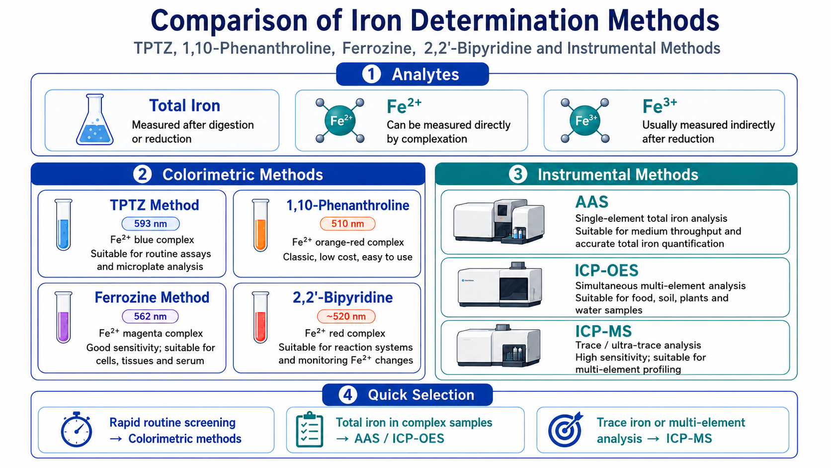
Iron content detection is used to evaluate total iron, ferrous iron, ferric iron, or bioavailable iron levels in water samples, foods, soils, plants, cells, serum, tissues, and fermentation systems. Common methods can be divided into colorimetric assays and instrumental analytical methods. TPTZ, 1,10-phenanthroline, Ferrozine, and 2,2'-bipyridine methods are all based on Fe²⁺ complexation and color development, making them suitable for routine detection and iron metabolism studies. AAS, ICP-OES, and ICP-MS are more suitable for total iron, multi-element, and trace iron quantification.
Keywords: iron content detection; total iron detection; ferrous iron detection; ferric iron detection; TPTZ method; phenanthroline method; Ferrozine method; 2,2'-bipyridine method; ICP-MS
1 Basic Logic of Iron Content Detection
1.1 Distinguishing detection targets
(1) Total iron
Total iron refers to the total amount of all iron species in a sample, including Fe²⁺, Fe³⁺, protein-bound iron, complexed iron, particulate iron, and mineral-bound iron. Total iron detection usually requires acid digestion, wet ashing, microwave digestion, or strong acid extraction to convert iron in different binding states into measurable forms.
(2) Ferrous iron
Ferrous iron mainly refers to Fe²⁺. It has relatively strong reducing properties and is easily oxidized to Fe³⁺ by air, peroxides, or high-pH environments. If the experimental goal is Fe²⁺ detection, samples should be processed as soon as possible after collection, with temperature, light exposure, acidification, and oxygen exposure carefully controlled.
(3) Ferric iron
Fe³⁺ often exists as complexes, hydroxides, protein-bound forms, or precipitates. Most colorimetric methods do not directly detect Fe³⁺. Instead, Fe³⁺ is first reduced to Fe²⁺, and then TPTZ, 1,10-phenanthroline, Ferrozine, or 2,2'-bipyridine is used to form colored complexes for measurement.
(4) Extractable iron and bioavailable iron
In plant, soil, and food samples, total iron, acid-extractable iron, chelated iron, and bioavailable iron are often distinguished. Different extraction systems generate results with different meanings, and “extractable iron” should not be directly equated with total iron in the sample.
Table 1 Detection targets and interpretation of iron content results
Detection target | Main meaning | Common samples | Method selection focus |
Total iron | Total amount of all iron species in the sample | Water, food, soil, tissue, cells, plants | Requires digestion, acid extraction, or reduction |
Fe²⁺ | Reduced ferrous iron content | Water, culture medium, cell extracts, reducing systems | Oxidation must be prevented; suitable for Fe²⁺ complexation colorimetric assays |
Fe³⁺ | Oxidized ferric iron content | Water, serum, tissue extracts, culture systems | Usually indirectly detected after reduction |
Extractable iron | Iron released by a specific extractant | Soil, plants, food | Results are affected by extractant and pH |
Bioavailable iron | Iron available to cells or biological systems | Food, culture systems, nutrition studies | Requires analysis combined with in vitro digestion, cell models, or chelation status |
1.2 Key factors in method selection
(1) Detection purpose
If only rapid comparison of iron content between groups is required, colorimetric methods such as TPTZ, 1,10-phenanthroline, Ferrozine, or 2,2'-bipyridine can be selected. If accurate total iron determination is required, especially for food, soil, plant, or tissue samples, AAS, ICP-OES, or ICP-MS after digestion should be prioritized.
(2) Iron speciation requirements
Direct color development usually reflects Fe²⁺ or iron that can be converted into Fe²⁺. If Fe²⁺ and Fe³⁺ need to be distinguished, an “unreduced group” and a “total iron reduced group” should be set up, with Fe³⁺ estimated by difference. If stricter speciation analysis is required, separation-coupled methods or dedicated iron speciation methods should be used.
(3) Sample matrix complexity
Serum, tissue, food, and soil samples have complex matrices. Proteins, organic compounds, salts, pigments, and particulates may affect color development or instrumental injection. Complex samples require deproteinization, filtration, digestion, dilution, or matrix matching.
(4) Detection throughput
Colorimetric assays are suitable for high-throughput microplate detection. AAS is suitable for batch single-element detection. ICP-OES and ICP-MS are suitable for simultaneous multi-element analysis, especially for combined detection of iron, zinc, copper, manganese, calcium, magnesium, and other elements.
2 Iron Colorimetric Assays
2.1 TPTZ method
(1) Detection principle
TPTZ, or 2,4,6-tris(2-pyridyl)-1,3,5-triazine, forms a blue Fe²⁺-TPTZ complex with Fe²⁺, which is commonly measured at approximately 593 nm. If used for total iron detection, Fe³⁺ is usually reduced to Fe²⁺ before complexation and color development.
(2) Application scenarios
The TPTZ method is suitable for iron content detection in water samples, food extracts, plant samples, serum, or some biological samples. It is also commonly used in iron-reducing capacity and FRAP-related systems. When used for iron content detection, it should be clarified whether the assay measures “iron complex color development” or “reducing capacity evaluation,” to avoid confusing antioxidant reducing capacity results with iron content.
(3) Method features
The TPTZ method has high color sensitivity and obvious color contrast, making it suitable for microplate detection. The reaction is usually performed under acidic conditions. Reducing substances, strong chelators, pigments, and turbidity in the sample may affect results. If the sample itself has strong reducing capacity, it may influence the conversion of Fe³⁺ to Fe²⁺, and sample blanks and spike recovery should be included.
2.2 1,10-Phenanthroline method
(1) Detection principle
1,10-Phenanthroline forms an orange-red complex with Fe²⁺, which is commonly measured at approximately 510 nm. If total iron is detected, hydroxylamine hydrochloride, ascorbic acid, or another reducing agent is added to reduce Fe³⁺ to Fe²⁺ before color development.
(2) Application scenarios
The phenanthroline method is suitable for water samples, environmental samples, culture media, and some pretreated food or biological samples. It is simple, low-cost, and is a classic method for routine colorimetric detection of total iron and ferrous iron.
(3) Method features
The phenanthroline method is suitable for routine laboratory colorimetric detection, but its anti-interference capability is limited. Copper, nickel, cobalt, and other metal ions, strong oxidants, strong reducing agents, chelators, and sample background color may all affect color development. For complex matrix samples, sample blanks, standard addition, and spike recovery should be used.
2.3 Ferrozine method
(1) Detection principle
Ferrozine forms a stable purple-red complex with Fe²⁺, which is commonly measured at approximately 562 nm. If Fe²⁺ is detected directly, no reducing agent is required. If total iron is detected, Fe³⁺ must first be reduced to Fe²⁺.
(2) Application scenarios
The Ferrozine method is commonly used for cells, tissues, serum, culture media, food extracts, and iron metabolism studies. Because it is sensitive to Fe²⁺ color development, it is often used in iron uptake, iron release, iron-reducing capacity, iron homeostasis, and ferroptosis-related iron content assays.
(3) Method features
The Ferrozine method has good sensitivity and is suitable for micro-samples and microplate assays. Results are affected by sample redox status, completeness of protein precipitation, completeness of iron release, competitive binding by chelators, and background color. For biological samples, acid extraction or protein precipitation should be carefully optimized.
2.4 2,2'-Bipyridine method
(1) Detection principle
2,2'-Bipyridine forms a colored complex with Fe²⁺, usually red to orange-red, and absorbance is typically measured around 520 nm. Similar to the phenanthroline method, if total iron is detected, Fe³⁺ must first be reduced to Fe²⁺.
(2) Application scenarios
The 2,2'-bipyridine method can be used for water samples, chemical reaction systems, reduction process evaluation, and some metal complexation studies. It can also be used for colorimetric determination of serum iron and other specific samples. This method is suitable for observing Fe²⁺ formation, Fe³⁺ reduction, and iron ion complexation reactions.
(3) Method features
The 2,2'-bipyridine method is visually straightforward and suitable for Fe²⁺-related chemical system analysis. Its limitation is relatively weak selectivity and anti-interference ability. Other transition metal ions, chelators, reducing agents, and color backgrounds in samples may affect results. Method validation is required when used for complex samples.
Table 2 Comparison of Four Fe²⁺ Complexation Colorimetric Methods
Method | Chromogenic Reagent | Main Detection Target | Typical Color | Common Detection Wavelength | Main Applications |
TPTZ method | TPTZ | Fe²⁺; total iron after reduction | Blue | Approx. 593 nm | Total iron colorimetry, iron reduction systems, microplate detection |
1,10-Phenanthroline method | 1,10-Phenanthroline | Fe²⁺; total iron after reduction | Orange-red | Approx. 510 nm | Water samples, environmental samples, routine total iron detection |
Ferrozine method | Ferrozine | Fe²⁺; total iron after reduction | Purple-red | Approx. 562 nm | Cells, tissues, serum, and iron metabolism studies |
2,2'-Bipyridine method | 2,2'-Bipyridine | Fe²⁺; total iron after reduction | Red to orange-red | Approx. 520 nm | Fe²⁺ complexation, serum iron detection, reduction processes, and chemical system analysis |
2.5 Prussian blue-related methods
(1) Detection basis
The Prussian blue reaction is mainly used to display Fe³⁺-related iron deposits. In histology, Perls’ Prussian blue staining can reveal ferric iron deposits such as hemosiderin. In solution systems, iron ions can form blue complexes with ferricyanide or ferrocyanide under specific conditions and can be used for iron detection in certain settings.
(2) Application scenarios
Prussian blue staining is more suitable for tissue localization than high-precision solution-based total iron quantification. If the research objective is to observe the location and distribution of iron deposits in liver, spleen, bone marrow, or tissue sections, Prussian blue staining has clear value.
(3) Interpretation limitations
Positive Prussian blue staining in tissue indicates reactive ferric iron deposition and does not equal the total iron content of the sample. If quantification is required, image analysis, chemical extraction colorimetry, or ICP-based methods should be combined.
3 Instrumental Analysis Methods for Iron Content Detection
3.1 Atomic absorption spectroscopy
(1) Detection principle
Atomic absorption spectroscopy (AAS) quantifies iron by measuring the absorption intensity of characteristic wavelength light by ground-state iron atoms. Samples usually require acid digestion or acid extraction to convert iron into a solution state suitable for nebulization and atomization.
(2) Method types
Flame atomic absorption is suitable for samples with relatively high iron content, while graphite furnace atomic absorption has higher sensitivity and is suitable for micro- or trace-level iron detection. Method selection should be based on sample concentration range, matrix complexity, and detection throughput.
(3) Technical features
AAS has good selectivity, mature methodology, and lower cost than ICP-MS, but it is usually mainly used for single-element detection. If only iron needs to be measured and sample numbers are moderate, AAS is highly practical. If multiple elements need to be detected simultaneously, ICP-OES or ICP-MS is more suitable.
3.2 ICP-OES
(1) Detection principle
ICP-OES uses inductively coupled plasma to excite elements in the sample, causing them to emit characteristic spectra. Quantification is performed based on emission intensity. This method is suitable for simultaneous multi-element detection and is commonly used for total iron analysis in foods, soils, plants, water samples, and biological samples.
(2) Application scope
ICP-OES is suitable for medium-low to relatively high iron content detection, with a wide linear range and high throughput. For samples where iron content is not extremely low and other mineral elements need to be detected simultaneously, ICP-OES often offers good cost-effectiveness.
(3) Technical features
This method requires high-quality sample digestion, and salt content, acidity, matrix effects, and spectral interferences must be controlled. Complex samples should use internal standards, matrix matching, dilution, or standard addition to improve accuracy.
3.3 ICP-MS
(1) Detection principle
ICP-MS uses plasma to ionize the sample, then separates and quantifies ions based on mass-to-charge ratio. This method has high sensitivity and is suitable for trace and ultra-trace iron detection, as well as multi-element combined analysis.
(2) Application scope
ICP-MS is suitable for low-iron samples, micro-tissue samples, low-volume samples, environmental trace contaminants, and metal metabolism analysis. If sample volume is limited and multi-element spectral information is required, ICP-MS has greater advantages.
(3) Technical features
ICP-MS is highly sensitive, but iron detection may be affected by polyatomic ion interference and matrix effects. Appropriate isotopes, collision/reaction cell conditions, internal standard systems, and digestion workflows are required. Instrument cost and maintenance cost are high, making it unsuitable for simple routine screening.
Table 3 Comparison of AAS, ICP-OES, and ICP-MS
Method | Detection Target | Sensitivity | Throughput | Main Advantages | Main Limitations |
Flame AAS | Total iron | Moderate | Moderate | Mature method with relatively low cost | Limited multi-element capability and sensitivity |
Graphite Furnace AAS | Total iron / trace iron | High | Relatively low | Suitable for trace iron detection | Low throughput and high requirements for matrix control |
ICP-OES | Total iron and multiple elements | Medium to high | High | Simultaneous multi-element detection and wide linear range | Requires strict control of digestion quality and spectral interferences |
ICP-MS | Trace iron and multiple elements | Very high | High | Ultra-trace detection with strong multi-element capability | High cost and requires control of mass spectral interferences |
4 Comparison of Sample Pretreatment Methods
4.1 Water samples and culture media
(1) Filtration and acidification
Water samples and culture media usually require filtration to remove particulates and acid preservation to reduce iron precipitation and container adsorption. If dissolved iron is studied, samples should be filtered as soon as possible after collection. If total iron is studied, particulate iron should be retained and digested.
(2) Fe²⁺ protection
Ferrous iron detection is highly sensitive to oxidation. Samples should be tested as soon as possible. Acidification, protection from light, low temperature, and nitrogen protection may be used when necessary. If detection is delayed, Fe²⁺ may be converted to Fe³⁺, causing underestimation of ferrous iron.
(3) Matrix blank
Phosphates, peptones, metal salts, and chelators in culture media may contribute background iron or interfere with color development. Medium blanks and spike recovery should be included.
4.2 Biological samples
(1) Serum and plasma
Serum iron detection often involves distinguishing transferrin-bound iron, free iron, and total iron. If colorimetric methods are used, hemolysis interference should be considered, because hemoglobin iron released from red blood cells can significantly affect results.
(2) Cells and tissues
Total iron detection in cells and tissues usually requires lysis, acid hydrolysis, or wet digestion. If the Ferrozine, TPTZ, or phenanthroline method is used, sufficient protein precipitation, complete iron release, and sample blanks should be ensured.
(3) Plant samples
Iron in plant tissues is often bound to cell walls, chloroplasts, proteins, and organic acids. Total iron analysis is often performed by ICP-OES, ICP-MS, or AAS after acid digestion. If extractable iron is detected, the extractant, pH, and extraction time must be clearly defined.
4.3 Food, soil, and complex matrices
(1) Food samples
Proteins, polyphenols, phytic acid, fats, and pigments in foods may interfere with colorimetric methods. Total iron detection often uses wet digestion or microwave digestion followed by AAS or ICP analysis.
(2) Soil samples
Iron species in soil are complex and include mineral-bound, complexed, exchangeable, and reducible iron. Extraction methods for total iron and available iron differ, and the results cannot be substituted for one another.
(3) Complex matrix control
Complex samples should use reference materials, spike recovery, method blanks, replicate samples, and matrix-matched standard curves to control accuracy. Using only water-based standard curves may produce significant matrix bias.
Table 4 Pretreatment strategies for different sample types
Sample type | Recommended pretreatment | Suitable methods | Notes |
Water sample | Filtration, acidification, or digestion | TPTZ method, phenanthroline method, AAS, ICP | Distinguish dissolved iron from total iron |
Culture medium | Centrifugation, filtration, matrix blank | Ferrozine method, TPTZ method, ICP-OES | Medium background iron must be subtracted |
Serum/plasma | Avoid hemolysis; deproteinize if necessary | 2,2'-Bipyridine method, Ferrozine method, AAS, ICP-MS | Hemolysis significantly increases results |
Cells | Lysis, acid hydrolysis, protein precipitation | Ferrozine method, TPTZ method, ICP-MS | Normalize by cell number or protein content |
Tissue | Homogenization, acid digestion, or wet digestion | Ferrozine method, non-heme iron detection, AAS, ICP-OES | Ensure complete iron release |
Plants | Drying, grinding, acid digestion | ICP-OES, AAS | Avoid external soil contamination |
Food | Homogenization, ashing, or acid digestion | AAS, ICP-OES, ICP-MS, non-heme iron detection | Pigments and organic matter affect colorimetry |
Soil | Acid digestion or extractant extraction | Phenanthroline method, ICP-OES, AAS | Total iron and available iron should not be mixed |
5 Method Selection and Result Interpretation
5.1 Selecting methods by experimental purpose
(1) Routine total iron screening
If the sample matrix is simple and iron content is relatively high, the phenanthroline method, Ferrozine total iron method, TPTZ method, or 2,2'-bipyridine method can be selected. These methods are low-cost, fast, and suitable for preliminary screening and batch comparison.
(2) Iron metabolism research in biological samples
For cells, tissues, and serum samples, the Ferrozine method is widely used. The TPTZ method can be used for total iron colorimetry or reduction system-related assays. Phenanthroline and 2,2'-bipyridine are more suitable for routine Fe²⁺ complexation colorimetry or chemical system analysis. If higher accuracy or multi-element information is required, ICP-OES or ICP-MS should be used.
(3) Trace iron or multi-element analysis
Low-abundance iron detection, micro-samples, and multi-element combined analysis are more suitable for ICP-MS. If iron content is relatively high and multiple metal elements need to be detected simultaneously, ICP-OES is usually more suitable.
(4) Tissue iron deposition localization
If the goal is to observe iron deposition location rather than solution-based quantification, Prussian blue staining or enhanced tissue iron staining should be selected and combined with image analysis or pathological scoring.
Table 5 Selection of iron content detection methods
Experimental objective | Recommended methods | Reason for selection | Notes |
Simple water sample total iron detection | Phenanthroline method, TPTZ method, Ferrozine method | Simple operation, low cost | Requires sample blank and reduction step |
Fe²⁺ detection | Ferrozine method, TPTZ method, phenanthroline method, 2,2'-bipyridine method | Clear Fe²⁺ complex color development | Prevent Fe²⁺ oxidation |
Iron-reducing capacity-related detection | TPTZ method, Ferrozine method | Sensitive to Fe²⁺ formation | Distinguish iron content from reducing capacity |
Cellular total iron detection | TPTZ method, Ferrozine method, ICP-MS | Suitable for cell iron metabolism, ferroptosis, and oxidative stress studies | Normalize by protein content, cell number, or DNA amount |
Serum iron detection | 2,2'-Bipyridine method, AAS, ICP-MS | Suitable for serum iron content analysis | Hemolyzed samples should be excluded or separately labeled |
Non-heme iron detection | Bathophenanthroline method, Ferrozine method | Suitable for food, tissue, or biological samples | Distinguish heme iron from non-heme iron |
Food iron content | AAS, ICP-OES, ICP-MS | Higher accuracy, suitable for digested samples | Control digestion blanks and reference materials |
Soil total iron | Phenanthroline method, ICP-OES, AAS | Suitable for soil total iron or extractable iron detection | Distinguish from available iron detection |
Tissue iron deposition localization | Prussian blue staining | Displays iron deposition location | Not equivalent to total iron quantification |
5.2 Common abnormal results
(1) Colorimetric results are too high
Possible causes include sample color, turbidity, reducing agent background, metal ion interference, or container contamination. Sample blanks, reagent blanks, and spike recovery should be included, and low-metal-background consumables should be used.
(2) Colorimetric results are too low
Possible causes include incomplete iron release, Fe²⁺ oxidation, competitive binding by chelators, or incomplete protein precipitation. In total iron detection, the reduction step and digestion/extraction step should be checked for completeness.
(3) ICP result fluctuation
Possible causes include incomplete digestion, excessive matrix salt, contamination of the sample introduction system, spectral interference, or internal standard drift. Quality control samples, internal standards, blanks, and replicate measurements should be used for troubleshooting.
Table 6 Common interferences and controls in iron content detection
Interference source | Effect | Mainly affected methods | Control strategy |
Sample color | Increases background absorbance | Colorimetric methods | Set sample blank; dilute or purify if necessary |
Turbidity/particulates | Light scattering causes high absorbance | Colorimetric methods | Centrifuge, filter, or digest |
Hemolysis | Hemoglobin iron release | Serum/plasma detection | Avoid hemolyzed samples; record sample status |
Strong chelators | Prevent iron binding with chromogenic reagent | Colorimetric methods | Optimize pretreatment or detect after digestion |
Redox changes | Changes Fe²⁺/Fe³⁺ ratio | Speciation detection | Rapid detection, acidification, light protection, low temperature |
Metal contamination | Increases blank value | All methods | Use acid-washed consumables and ultrapure water |
Matrix salts | Nebulization or ionization interference | AAS, ICP | Dilution, matrix matching, internal standard correction |
Incomplete digestion | Insufficient iron release | AAS, ICP | Optimize acid system and digestion program |
6 Reagents and Materials Related to Iron Content Detection
Table 7 Selection of key reagents and materials for iron content detection
Product/Material Name | CAS No. | Product Category | Application Positioning |
TPTZ | Iron chromogenic reagent | Used for blue Fe²⁺-TPTZ complexation, total iron detection after reduction, and iron-reducing capacity-related experiments | |
1,10-Phenanthroline | Iron chromogenic reagent | Used for orange-red Fe²⁺ complexation and total iron colorimetric detection after reduction | |
Ferrozine | Ferrous iron chromogenic reagent | Used for Fe²⁺ detection, cellular/tissue iron content detection, and iron-reducing capacity analysis | |
2,2'-Bipyridine | Ferrous iron complexation chromogenic reagent | Used for Fe²⁺ complexation color development, Fe³⁺ reduction process analysis, and chemical system iron detection | |
Bathocuproine disulfonic acid disodium salt (BPS) | Iron complexation chromogenic reagent | Used for Fe²⁺ complexation detection and iron metabolism-related experiments | |
Hydroxylamine hydrochloride | Reducing agent | Used to reduce Fe³⁺ to Fe²⁺ for total iron colorimetric detection | |
Ascorbic acid | Reducing agent | Used for iron reduction treatment and Fe³⁺ to Fe²⁺ conversion | |
Sodium acetate | Buffer salt | Used to adjust color development pH in phenanthroline, Ferrozine, or TPTZ methods |
Table 8 Product selection for iron content detection, iron standards, and tissue iron deposition localization
Cat. No. | Product Name | Grade/Specification | Product Category | Application Positioning |
Soil Total Iron Content Assay Kit (o-Phenanthroline, Micro Method) | BioReagent | Soil total iron assay kit | Used for micro-method detection of total iron in soil samples; suitable for batch screening using the phenanthroline system | |
Soil Total Iron Content Assay Kit (Phenanthroline, Colorimetric Method) | BioReagent | Soil total iron assay kit | Used for colorimetric detection of total iron in soil samples; suitable for routine spectrophotometric analysis | |
Cell Total Iron Ion Content Assay Kit (TPTZ, Micro Method) | BioReagent | Cellular total iron assay kit | Used for total iron detection in cell samples; suitable for TPTZ chromogenic systems and microplate reading | |
Total Iron Ion Content Assay Kit (TPTZ, Micro Method) | BioReagent | Total iron assay kit | Used for total iron ion detection in samples; suitable for Fe³⁺ reduction followed by TPTZ complexation colorimetric analysis | |
Serum Iron Content Assay Kit (2, 2‘-Bipyridine, Micro Method) | BioReagent | Serum iron assay kit | Used for micro-method detection of iron content in serum samples; suitable for 2,2'-bipyridine colorimetric systems | |
Serum Iron Content Assay Kit (2, 2’-Bipyridine, Colorimetric Method) | BioReagent | Serum iron assay kit | Used for serum iron colorimetric detection; suitable for routine spectrophotometric workflows | |
Non-Heme Iron Content Assay Kit (Bathophenanthroline, Micro Method) | BioReagent | Non-heme iron assay kit | Used for micro-method detection of non-heme iron in food, tissue, or biological samples | |
Non-Heme Iron Content Assay Kit (Bathophenanthroline, Colorimetric Method) | BioReagent | Non-heme iron assay kit | Used for colorimetric detection of non-heme iron; suitable for nutrition, food, and tissue iron analysis | |
Iron Content Assay Kit (Ferrozine, Micro Method) | BioReagent | Iron content assay kit | Used for micro-method iron content detection; suitable for Ferrozine ferrous iron complexation colorimetric systems | |
Iron Content Assay Kit (Ferrozine, Colorimetric Method) | BioReagent | Iron content assay kit | Used for iron content colorimetric detection; suitable for routine Ferrozine spectrophotometric analysis | |
Ferrous Ion Content Assay Kit (TPTZ, Micro Method) | BioReagent | Ferrous iron assay kit | Used for micro-method Fe²⁺ detection; suitable for TPTZ complexation color development and microplate reading | |
Ferrous Ion Content Assay Kit (TPTZ, Colorimetric Method) | BioReagent | Ferrous iron assay kit | Used for colorimetric Fe²⁺ detection; suitable for spectrophotometric analysis | |
Ferrous Ion Content Assay Kit (Ferrozine, Micro Method) | BioReagent | Ferrous iron assay kit | Used for micro-method Fe²⁺ detection; suitable for Ferrozine-based ferrous iron analysis | |
Ferrous Ion Content Assay Kit (Ferrozine, Colorimetric Method) | BioReagent | Ferrous iron assay kit | Used for colorimetric Fe²⁺ detection; suitable for ferrous iron analysis in water samples, culture media, or biological extracts | |
Cell Ferrous Ion Content Assay Kit (TPTZ, Micro Method) | BioReagent | Cellular ferrous iron assay kit | Used for Fe²⁺ detection in cell samples; suitable for iron metabolism, redox, and ferroptosis-related experiments | |
Ferrous sulfate standard solution | 0.05mol/L in H2O | Fe²⁺ standard solution | Used for low-concentration ferrous iron standard curves, method validation, and QC sample preparation | |
FERROUS SULFATE standard solution | 0.1mol/L in H2O | Fe²⁺ standard solution | Used for Fe²⁺ colorimetric standard curves and ferrous iron recovery experiments | |
FERROUS SULFATE standard solution | 0.2mol/L in H2O | Fe²⁺ standard solution | Used for ferrous iron method validation, chromogenic system calibration, and QC | |
FERROUS SULFATE standard solution | 0.25mol/L in H2O | Fe²⁺ standard solution | Used as a relatively high-concentration Fe²⁺ standard stock and for method validation | |
Ferrous Sulfate standard solution | 0.5mol/L in H2O | Fe²⁺ standard solution | Used as a ferrous standard stock solution, for gradient dilution, and assay system calibration | |
FERROUS SULFATE standard solution | 1mol/L in H2O | Fe²⁺ standard solution | Used as a high-concentration Fe²⁺ standard stock and for preparing wide-range standard curves | |
Iron Standard | 100μg/mL | Iron element standard solution | Used for preparing low-concentration iron standard curves in colorimetry, AAS, or ICP | |
Iron Standard | 100μg/ml in 5%HCl | Iron element standard solution | Used for iron standard curves, spike recovery, and QC in acidic matrices | |
Iron standard | 500mg/L in 1%HCl | Iron element standard solution | Used as a medium-high concentration iron standard stock in instrumental analysis or colorimetric detection | |
Iron resolution | analytical standard, 1000ug/ml in 1.0mol/L HNO3 | Iron element standard solution | Used for iron standard curves in AAS, ICP-OES, ICP-MS, and method validation | |
Iron solution | analytical standard, 1000mg/L in 5%HCl | Iron element standard solution | Used for iron content standard curves, spike recovery, and instrument calibration | |
Prussian blue | Biological Stain | Tissue iron staining dye | Used for ferric iron deposition visualization in tissue sections and preparation of Prussian blue staining systems | |
Prussian blue soluble | Biological Stain | Tissue iron staining dye | Used for Prussian blue-related staining or iron deposition visualization systems | |
Prussian Blue Staining Solution (DAB Enhancement Method) | BioReagent,for microscopy,Biological Stain | Tissue iron staining solution | Used for tissue iron deposition localization; DAB enhancement helps improve weak positive iron deposition signals | |
Prussian Blue Staining Kit (Neutral Red) | BioReagent, Biological Stain, for microscopy | Tissue iron staining kit | Used for ferric iron deposition localization in tissue sections, with neutral red counterstaining for background structure | |
Prussian Blue Staining Kit (Eosin) | BioReagent, Biological Stain, for microscopy | Tissue iron staining kit | Used for tissue iron deposition localization; suitable for observing tissue background structure with eosin counterstaining | |
Prussian Blue Staining Kit (Nuclear Fast Red) | BioReagent, for microscopy, Biological Stain | Tissue iron staining kit | Used for tissue section iron deposition localization; nuclear fast red counterstaining supports nuclear and tissue structure interpretation |
7 Quality Control and Method Validation
7.1 Standard curve and linear range
(1) Standard curve
Colorimetric methods, AAS, and ICP methods all require standard curves. Standard points should cover the expected concentration range of the samples, and sample absorbance or signal intensity should not remain near the edge of the linear range.
(2) Blank control
Reagent blanks are used to subtract reagent background. Sample blanks are used to subtract sample color and turbidity. Method blanks are used to evaluate exogenous iron contamination introduced by digestion, containers, and water systems.
(3) Spike recovery
Spike recovery should be performed for complex matrix samples. Abnormal recovery indicates matrix interference, incomplete color development, incomplete digestion, or instrumental matrix effects.
7.2 Result normalization
(1) Cell samples
Cellular iron content can be normalized by cell number, protein content, or DNA content. Different normalization methods reflect different biological meanings, and the method should be fixed at the experimental design stage.
(2) Tissue samples
Tissue iron can be calculated by wet weight, dry weight, or protein content. Animal and plant tissues commonly use dry weight or wet weight, while cellular biochemical experiments often use protein content.
(3) Environmental and food samples
Water samples are commonly expressed as mg/L or μg/L, while food and soil samples are often expressed as mg/kg. When different units are used, sample dilution, digestion, and wet/dry weight conversion should be clearly stated.
8 Common Questions
8.1 Does the TPTZ method detect iron content or reducing capacity?
TPTZ can be used for Fe²⁺ complexation color development and is also commonly used in FRAP and other reducing capacity assays. If used for iron content detection, an iron standard curve should be established and the reduction step controlled. If used for FRAP, the result mainly reflects the sample’s ability to reduce Fe³⁺ to Fe²⁺ and should not be directly equated with iron content.
8.2 Does the Ferrozine method detect total iron or ferrous iron?
Ferrozine itself mainly detects Fe²⁺. If Ferrozine is added directly, the result mainly reflects Fe²⁺ or iron that can immediately form Fe²⁺ complexes in the system. If Fe³⁺ is first reduced to Fe²⁺ using a reducing agent, it can be used for total iron detection.
8.3 What is the difference between the phenanthroline method and the 2,2'-bipyridine method?
Both can form colored complexes with Fe²⁺. The phenanthroline method is more commonly used for water samples, soil extracts, and routine total iron colorimetric detection, and the method is well established. The 2,2'-bipyridine method can be used for serum iron detection, Fe²⁺ complexation, iron reduction processes, and chemical system analysis. In complex biological samples, the Ferrozine and TPTZ methods are also commonly used.
8.4 Should iron content detection use colorimetry or ICP?
If the sample matrix is simple and the detection goal is routine screening or group comparison, colorimetry is usually sufficient. If high accuracy, multi-element simultaneous analysis, trace detection, or complex matrix samples are involved, ICP-OES or ICP-MS is more suitable.
8.5 Why are colorimetric and ICP results inconsistent for the same sample?
The difference may come from pretreatment. Colorimetric methods detect iron that can be released and participate in the color reaction, whereas ICP usually detects total iron after digestion. If digestion completeness, reduction steps, complexation interference, or matrix background differ, results may not be consistent.
8.6 What is most important when detecting Fe²⁺?
Fe²⁺ is easily oxidized to Fe³⁺. Samples should be detected as soon as possible after collection, while controlling pH, temperature, air exposure, and light. Acidification, light protection, low temperature, and inert gas protection may be used when necessary to avoid underestimating ferrous iron.
Selection of iron content detection methods should be based on whether the goal is total content or speciation, simple or complex matrix, routine screening or trace quantification, and bulk content or tissue localization. TPTZ, 1,10-phenanthroline, Ferrozine, and 2,2'-bipyridine all rely on Fe²⁺ complexation and color development, but their suitable matrices and interference characteristics differ. AAS, ICP-OES, and ICP-MS are more suitable for accurate total iron detection and multi-element analysis, while Prussian blue staining is used for spatial localization of tissue iron deposition.