
ADC Linkers: Key Units That Regulate the In Vivo Stability, Payload Release, and Efficacy and Safety of Antibody–Drug Conjugates
ADC Linkers: Key Units That Regulate the In Vivo Stability, Payload Release, and Efficacy and Safety of Antibody–Drug Conjugates
1. Why Should ADCs Not Be Evaluated Based Only on the Antibody and the Payload?
Antibody–drug conjugates (ADCs) consist of three components: a monoclonal antibody, a linker, and a cytotoxic payload. The antibody is responsible for recognizing tumor-associated antigens, the payload is responsible for killing cells, and the linker determines whether the payload can be safely carried in vivo and released at the appropriate site.
The first point to understand is a fundamental trade-off:
The payload must be potent enough to kill tumor cells; however, it must not be released prematurely in the bloodstream, otherwise it may cause systemic toxicity.
Therefore, ADCs should not be evaluated only by whether the antibody is target-specific or whether the payload is highly potent. Even if the antibody has high affinity and the payload is strongly cytotoxic, premature linker cleavage in plasma can cause the ADC to lose its targeted delivery advantage and instead become a source of systemic exposure to a free toxic small molecule. Conversely, if the linker is overly stable, insufficient payload release after entry into the target cell may also reduce efficacy. The therapeutic effect of an ADC depends on the coordinated performance of all three components:
Component | Main Function | Key Questions |
Antibody | Recognizes tumor-associated antigens | Is the target specific? Can it mediate internalization? Is it expressed in normal tissues? |
Linker | Connects the antibody and payload, and controls release | Is it stable in blood? Can it release the payload inside target cells? |
Payload | Kills tumor cells | Is the cytotoxicity sufficient? Can the released payload reach its site of action? |
2. What Is an ADC Linker: From a “Connecting Structure” to a “Release-Control Unit”
An ADC linker is the covalent connecting unit between the antibody and the payload. One end is attached to the antibody, and the other end is attached to the payload. On the surface, the role of the linker is to “connect”; however, from the perspective of drug design, its true role is to control the in vivo release behavior of the payload.
An ideal linker needs to meet three requirements simultaneously.
2.1 Stable in Blood Circulation
After administration, an ADC needs to circulate in plasma and reach tumor tissue. If the linker breaks prematurely during circulation, the payload will be released too early, increasing exposure in normal tissues and reducing the effective ADC concentration that reaches tumor cells.
2.2 Effective Release Inside Target Cells
Many ADCs rely on antigen-mediated internalization to enter cells and then traffic into endosomes and lysosomes. The linker must take advantage of these intracellular environmental features to release the payload, such as low pH, proteases, highly reducing conditions, or lysosomal protein degradation processes.
2.3 No Significant Disruption of the Physicochemical Properties of the ADC
The linker–payload can alter the hydrophobicity, charge distribution, aggregation tendency, and pharmacokinetics (PK) of an ADC. If the linker–payload is excessively hydrophobic, the ADC may become more prone to aggregation, increased nonspecific uptake, and accelerated plasma clearance.
To better understand the role of ADC linkers in ADC drugs, they can be examined within the complete mechanism of action. The ADC linker is involved throughout the entire process of in vivo delivery, target-cell uptake, payload release, and cell killing. The following figure summarizes, in a flowchart format, the core role of the linker in regulating ADC stability, release mode, and therapeutic window.
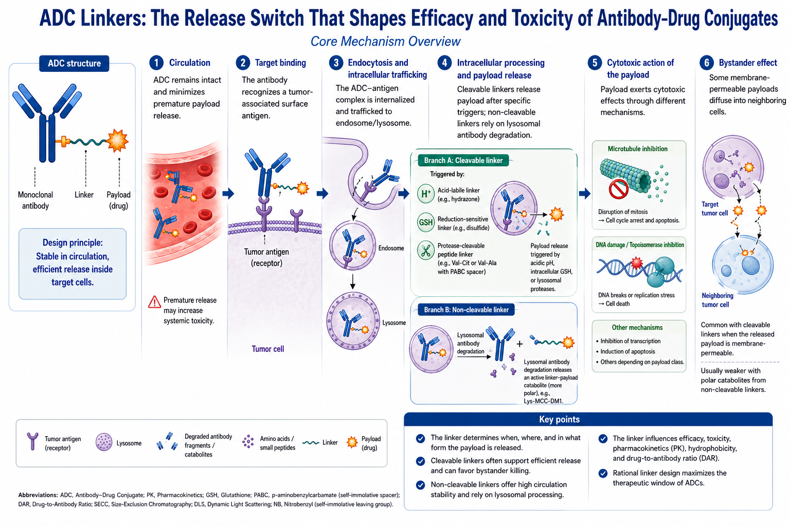
The figure above shows the main process by which an ADC undergoes blood circulation, target binding, internalization and intracellular trafficking, intracellular processing, payload release, and cell killing. Cleavable linkers can trigger payload release under acidic pH, intracellular glutathione (GSH), or lysosomal proteases; non-cleavable linkers usually rely on antibody degradation in lysosomes to release active metabolites carrying linker residues or amino acid residues.
After release, the payload can induce tumor cell death through mechanisms such as microtubule inhibition, DNA damage, or topoisomerase inhibition. When the payload has sufficient membrane permeability, it may also diffuse into neighboring cells and produce a bystander effect. The structural design of the linker simultaneously affects ADC efficacy, toxicity, pharmacokinetics, hydrophobicity, and drug-to-antibody ratio.
3. The Complete In Vivo Pathway of an ADC: Binding, Internalization, Release, and Cell Killing
From entering the body to killing tumor cells, a typical ADC usually undergoes the following steps.
3.1 Blood Circulation: The ADC Must Remain Intact
After entering the bloodstream, the antibody, linker, and payload must remain stable as an intact entity. The most important requirement at this stage is to prevent premature payload release. Insufficient plasma stability increases exposure to free payload and is one of the important sources of systemic toxicity associated with ADCs.
3.2 Target Binding: The Antibody Recognizes Antigens on the Tumor Cell Surface
The antibody binds to tumor cell surface antigens. Target selection should consider not only the expression level of the antigen in tumor tissue, but also its expression in normal tissues and whether antibody binding can induce internalization.
3.3 Internalization and Intracellular Trafficking: The ADC Enters Endosomes and Lysosomes
After the antibody–antigen complex is internalized, it enters early endosomes, then late endosomes, and eventually lysosomes. Lysosomes have an acidic environment and contain various proteases, making them key sites for payload release in many ADCs.
3.4 Linker Cleavage or Antibody Degradation: The Payload Forms an Active Molecule
For cleavable linkers, the payload can be released through acid-sensitive, reduction-sensitive, or enzyme-sensitive mechanisms. For non-cleavable linkers, the antibody usually needs to be degraded in lysosomes, releasing payload metabolites that carry the linker and amino acid residues.
3.5 Payload-Mediated Killing: Acting on Key Intracellular Targets
The released payload enters the cytoplasm or nucleus and acts on key targets such as microtubules, DNA, or topoisomerase I, thereby inducing cell-cycle arrest, DNA damage, or cell death.
3.6 Bystander Effect: Expanding the Killing Range in Antigen-Heterogeneous Tumors
If the released payload is membrane-permeable, it may diffuse from target antigen-positive cells into neighboring cells and kill cells with low or negative antigen expression. This phenomenon is known as the bystander effect. The bystander effect helps address heterogeneous tumor antigen expression, but it may also increase the risk of toxicity in normal tissues.
4. Cleavable Linkers: How Acidic, Reducing, and Enzymatic Environments Are Used to Release Payloads
The design concept of cleavable linkers is to use characteristic conditions inside target cells, especially in endosomes and lysosomes, or certain tumor-associated microenvironmental features, to cleave the linker at a specific site and release the payload. Common cleavable linkers include acid-sensitive linkers, reduction-sensitive linkers, and enzyme-sensitive linkers.
4.1 Acid-Sensitive Linkers: Using the Low pH of Endosomes and Lysosomes to Trigger Release
A common representative of acid-sensitive linkers is the hydrazone linker. Hydrazone bonds should ideally remain stable in the neutral plasma environment and undergo hydrolysis in acidic endosomal or lysosomal environments, thereby releasing the payload.
Simplified mechanism:
R¹R²C=N–NH–CO–Payload + H₂O + H⁺
→ R¹R²C=O + H₂N–NH–CO–Payload
The advantage of acid-sensitive linkers is that their trigger condition is clearly defined. Their main limitation is that plasma stability must be carefully controlled. If acid-sensitive bonds undergo nonspecific hydrolysis during circulation, premature payload release may occur.
4.2 Reduction-Sensitive Linkers: Using the Intracellular Reducing Environment to Trigger Release
A common representative of reduction-sensitive linkers is the disulfide linker. The intracellular concentration of reducing molecules such as glutathione (GSH) is usually higher than that in the extracellular environment, allowing disulfide bonds to be reduced and cleaved inside cells.
Simplified mechanism:
Antibody–Linker–S–S–Payload + 2 GSH
→ Antibody–Linker–SH + Payload–SH + GSSG
The release rate of disulfide linkers is influenced by neighboring groups around the disulfide bond, steric hindrance, and the intracellular reducing environment. The key design goal is to avoid premature reduction in the blood while ensuring efficient intracellular release.
4.3 Enzyme-Sensitive Linkers: Using Lysosomal Protease Cleavage
Enzyme-sensitive linkers are an important class in current ADC design. Typical structures include peptide linkers such as Val-Cit, Val-Ala, and GGFG. These linkers can be recognized and cleaved by lysosomal proteases, followed by payload release through a self-immolative spacer.
Among them, p-aminobenzyloxycarbonyl (PABC) is a common self-immolative spacer. Proteases first cleave the peptide segment, after which PABC undergoes a 1,6-elimination reaction to release the payload in a form close to the free payload. The specific release form depends on the payload attachment site and functional group type.
The following simplified mechanism uses an amine-containing payload linked through a carbamate bond as an example:
Antibody–Val–Cit–PABC–Payload
-- lysosomal protease cleavage →
Antibody–Val–Cit + H₂N–PABC–Payload
-- PABC self-immolative elimination →
Payload–NH₂ + p-quinone methide + CO₂
The advantage of enzyme-sensitive linkers is that the release process is highly aligned with the ADC internalization–lysosomal trafficking pathway. The main risk is that plasma enzyme activity differs across species, which may affect translational interpretation between preclinical models and humans.
5. Non-Cleavable Linkers: Why They Are More Stable but Usually Have Weaker Bystander Effects
Non-cleavable linkers do not rely on direct intracellular cleavage of a specific chemical bond. Instead, after the ADC is internalized by cells, the antibody portion is degraded by proteases in lysosomes, ultimately releasing payload metabolites that carry the linker and amino acid residues.
A typical example is T-DM1, or trastuzumab emtansine. T-DM1 uses the SMCC crosslinking reagent to form a non-cleavable MCC thioether linkage, thereby conjugating trastuzumab with DM1. After the ADC enters HER2 (Human Epidermal Growth Factor Receptor 2)-positive cells, the antibody portion is degraded in lysosomes, generating active metabolites such as Lys-MCC-DM1.
Schematic release of a non-cleavable linker:
Antibody–MCC–DM1
-- lysosomal protein degradation →
Lys–MCC–DM1
The main characteristics of non-cleavable linkers include:
Feature | Explanation |
Relatively high plasma stability | The payload is less likely to detach prematurely during circulation |
Release depends on internalization and lysosomal degradation | Target internalization capacity has a greater impact on efficacy |
Released products often carry amino acid residues | The metabolites are relatively polar and usually have lower membrane permeability |
Bystander effect is usually weaker | Killing is more biased toward antigen-positive cells |
The advantage of non-cleavable linkers is their greater stability, with a generally lower risk of premature payload release during circulation. Their limitation is a stronger dependence on target-cell internalization and lysosomal processing. For targets with uniform antigen expression and high internalization efficiency, non-cleavable linkers can provide good selectivity. For solid tumors with pronounced antigen heterogeneity, insufficient bystander effect may limit efficacy.
6. How Linkers Affect Efficacy, Toxicity, PK, DAR, and the Bystander Effect
The role of the linker is not limited to controlling release. It also systematically affects ADC efficacy, safety, and in vivo distribution.
6.1 Efficacy: Determining Whether the Payload Can Reach an Effective Concentration Inside Target Cells
ADC-mediated cell killing depends on the concentration of active payload released inside cells. If linker release is insufficient, the payload may fail to reach an effective concentration even after successful antibody binding and internalization. If release is too rapid or occurs too early, the effective amount delivered to the tumor site may be reduced.
6.2 Toxicity: Determining Systemic Exposure to Free Payload
Payloads are usually highly cytotoxic. If the linker is unstable in blood, systemic exposure to free payload or payload-related metabolites will increase. Clinical studies and systematic reviews suggest that linker cleavability, DAR, and systemic free-payload exposure may affect ADC systemic toxicity, but the specific risk must still be evaluated comprehensively based on ADC type, target, payload, and exposure level.
6.3 PK: Affecting ADC Clearance, Distribution, and Exposure
The hydrophobicity of the linker–payload can affect ADC aggregation tendency, nonspecific uptake, and plasma clearance. Studies have shown that increasing the drug-to-antibody ratio (DAR) can enhance in vitro cytotoxicity, but it may also accelerate plasma clearance due to increased overall hydrophobicity and thereby reduce in vivo exposure. This issue can be improved through hydrophilic linkers or PEGylated designs.
6.4 DAR: Affecting Potency, Stability, and Safety
DAR refers to the average number of payload molecules conjugated to each antibody. A DAR that is too low may result in insufficient killing potency, whereas a DAR that is too high may increase hydrophobicity, aggregation, clearance, and toxicity risk. The hydrophilicity of the linker, the conjugation method, and the conjugation site can affect DAR distribution, conjugation homogeneity, and whether the ADC can maintain acceptable physicochemical properties under different DAR conditions.
6.5 Bystander Effect: Affecting the Ability to Kill Antigen-Heterogeneous Tumors
When a cleavable linker releases a payload with good membrane permeability, a bystander effect is usually more likely to occur. In contrast, residual metabolite products released from non-cleavable linkers usually have lower membrane permeability and weaker bystander effects. Whether a bystander effect is desirable should be assessed based on tumor antigen-expression uniformity and the risk of toxicity in normal tissues.
7. How to Select an ADC Linker: Five Evaluation Dimensions
There is no fixed answer for ADC linker selection. The choice must be evaluated comprehensively according to the target, payload, tumor characteristics, and safety requirements.
7.1 Whether the Target Undergoes Efficient Internalization
If the target can rapidly internalize after antibody binding and enter the lysosomal pathway, enzyme-sensitive linkers or non-cleavable linkers may both be suitable. If target internalization efficiency is low, insufficient intracellular release may become a limiting factor, and the suitability of the target for ADC design needs to be re-evaluated.
7.2 Whether Antigen Expression Is Uniform
If tumor cells generally express high levels of the target antigen, designs with stronger selectivity and lower bystander effects may be prioritized. If tumor antigen expression is highly heterogeneous, cleavable linker–payload combinations with bystander effects may have an advantage.
7.3 Whether the Payload Needs to Act in a Free Form
Some payloads need to be released in a form close to the free payload to fully exert their activity. In such cases, a self-immolative spacer is very important. If the released product carries a large residual group, binding between the payload and its target may be affected.
7.4 Whether the Linker–Payload Is Excessively Hydrophobic
Highly hydrophobic linker–payloads tend to increase ADC aggregation and nonspecific clearance. For highly hydrophobic payloads or high-DAR ADCs, hydrophilic linkers, PEG spacers, or site-specific conjugation strategies should be considered to improve overall properties.
7.5 Whether the Safety Window Is Sufficient
If the target is also expressed to some extent in normal tissues, or if the payload is extremely cytotoxic, greater attention should be paid to plasma stability and release selectivity. If the therapeutic goal is to treat solid tumors with pronounced antigen heterogeneity, the need for a bystander effect should be evaluated under the premise that safety remains controllable.
8. How to Verify Whether a Linker Design Is Reasonable in Research
Whether a linker design is reasonable should be verified step by step through in vitro, cellular, and in vivo experiments.
8.1 Plasma Stability
The integrity, DAR changes, and free-payload release of an ADC after incubation in human plasma and relevant animal plasma should be measured. This experiment is used to determine whether the linker carries a risk of premature cleavage or deconjugation during circulation.
8.2 Conditional Release Experiments
Corresponding release experiments should be designed according to linker type:
Linker Type | Key Test Conditions | Evaluation Focus |
Acid-sensitive linker | Different pH conditions | Release under acidic conditions; stability under neutral conditions |
Disulfide linker | GSH or other reducing conditions | Release rate under reducing conditions |
Peptide linker | Cathepsins; lysosomal extracts | Cleavage efficiency and selectivity |
Non-cleavable linker | Cell internalization and lysosomal degradation models | Formation of active metabolites |
8.3 DAR and Conjugation Homogeneity Analysis
Liquid chromatography–mass spectrometry, hydrophobic interaction chromatography, and reduced and non-reduced mass spectrometry can be used to analyze DAR and conjugation distribution. The broader the DAR distribution, the more difficult it is to control ADC batch-to-batch consistency and in vivo behavior.
8.4 Aggregation and Physicochemical Stability
SEC-HPLC, dynamic light scattering, differential scanning fluorimetry, or differential scanning calorimetry can be used to evaluate ADC aggregation, purity, and thermal stability. Even if the release mechanism of a linker–payload is reasonable, it is not suitable for further development if it causes significant aggregation.
8.5 Comparison of Cytotoxicity in Antigen-Positive and Antigen-Negative Cells
An ADC should show stronger killing activity against antigen-positive cells and weaker killing activity against antigen-negative cells. This experiment is used to confirm whether ADC-mediated killing depends on antibody-targeted delivery rather than free payload or nonspecific uptake.
8.6 Bystander Effect Experiments
Antigen-positive and antigen-negative cells can be co-cultured to observe whether the ADC can kill neighboring antigen-negative cells. This experiment is suitable for evaluating the diffusion-mediated killing ability of cleavable linker and membrane-permeable payload combinations.
8.7 In Vivo PK, Efficacy, and Safety
In vivo evaluation should simultaneously measure total antibody, conjugated antibody, free payload, tumor inhibition rate, and toxicity indicators. Only by analyzing stability, release efficiency, tumor exposure, and normal-tissue toxicity within the same system can it be determined whether the linker truly improves the therapeutic window of an ADC.
9. Classification Tables of Representative Chemicals Related to ADC Linkers and Payloads
Table 1. Enzyme-Sensitive Cleavable Linkers and Self-Immolative Release Units
Category | CAS No. | Aladdin Cat. No. | Name | Specification or Purity | Product Features and Applications |
Tetrapeptide cleavable linker fragment | 1599440-20-6 | Gly-Gly-Phe-Gly-NH-CH2-O-CH2COOH | Moligand™, ≥98% | Contains a Gly-Gly-Phe-Gly protease-recognition sequence; used for the design of lysosomal enzyme-responsive linkers and payload-release studies | |
Val-Cit dipeptide linker core fragment | 159858-33-0 | Val-Cit | Moligand™, ≥98% | A classic cathepsin-sensitive dipeptide unit used to construct cleavable ADC linkers | |
Val-Cit-PAB protected intermediate | 159858-22-7 | Fmoc-Val-Cit-PAB-OH | ≥97% | Contains an Fmoc protecting group, a Val-Cit dipeptide, and a PAB self-immolative structure; used for stepwise synthesis of cleavable linkers | |
Val-Cit-PAB hydrophilic click intermediate | 2055041-40-0 | Azido-PEG1-Val-Cit-PAB-OH | ≥96% | Contains an azide group, a PEG1 spacer, and a Val-Cit-PAB structure; used to construct click-conjugation enzyme-sensitive linkers | |
Val-Cit-PABC activated linker | 159857-81-5 | Mc-Val-Cit-PABC-PNP | ≥97% | Contains a maleimide conjugation terminus, a Val-Cit-PABC release unit, and a PNP activating group; used to prepare cleavable linker–payload conjugates | |
Val-Cit-PAB maleimide linker | 159857-80-4 | N-[6-(2,5-dioxo-2,5-dihydro-1H-pyrrol-1-yl)hexanoyl]-L-valyl-N5-(carbamoyl)-[4-(hydroxymethyl)phenyl]-L-ornithinamide | ≥97% | Contains a maleimide conjugation terminus and a Val-Cit-PAB release structure; used in thiol-conjugated ADC linker research | |
Val-Cit-PAB activated release unit | 2210262-26-1 | Val-Cit-PAB-OSBT | —— | Contains a Val-Cit-PAB self-immolative release structure and an activated terminal group; used to prepare linker–payload conjugates | |
Val-Ala-PAB dipeptide release unit | 1343476-44-7 | Val-Ala-PAB | ≥99% | Contains a Val-Ala dipeptide and a PAB self-immolative structure; used for lysosomal protease-responsive release studies | |
Val-Ala-PAB maleimide linker | 1870916-87-2 | Mc-Val-Ala-PAB | ≥98% | Contains a maleimide conjugation terminus and a Val-Ala-PAB release structure; used to construct cysteine-conjugated ADC linkers | |
Val-Ala-PAB activated linker | 1639939-40-4 | MC-Val-Ala-PAB-PNP | Moligand™, ≥97% | Contains an MC conjugation terminus, a Val-Ala-PAB release unit, and a PNP activating group; used to prepare enzyme-sensitive linker–payload conjugates | |
VCP maleimide activated linker | 2395887-69-9 | Mal-PEG2-VCP-NB | Moligand™, ≥98% | Contains maleimide, a PEG2 spacer, and a VCP cleavable structure; used to construct thiol-conjugation and enzyme-responsive release systems |
Table 2. Acid-Sensitive, Reduction-Sensitive, and Hydrolytically Releasable Linkers
Category | CAS No. | Aladdin Cat. No. | Name | Specification or Purity | Product Features and Applications |
Acid-sensitive hydrazone linker precursor | 65623-82-7 | 4-(4-Acetylphenoxy)butyric acid | ≥95% | Contains an aromatic ketone structure; can be used to construct acid-sensitive hydrazone linkers and is suitable for release studies in acidic endosomal and lysosomal environments | |
Hydrolyzable SN38 linker unit | 2616704-22-2 | CL2A | ≥97% | Can be used in the design of hydrolytically releasable linkers for SN38-type payloads; suitable for ADC payload-release and stability studies | |
Reduction-sensitive disulfide crosslinking reagent | 68181-17-9 | SPDP (N-succinimidyl 3-(2-pyridyldithio)propionate) | ≥95% | Contains an NHS ester and a pyridyl disulfide group; used for amine modification, thiol exchange, and reduction-sensitive conjugation systems | |
Degradable disulfide linker | 115088-06-7 | SPDB, degradable ADC linker | ≥95% | Contains a sterically hindered disulfide structure; used for studies of reduction-triggered payload release | |
Water-soluble disulfide linker | 1193111-39-5 | sulfo-SPDB | Moligand™, ≥95% | Contains a sulfonate group and a disulfide structure; used to construct water-soluble, reduction-sensitive ADC linkers | |
PEGylated disulfide linker | 1305053-43-3 | PEG4-SPDP | Moligand™, ≥95% | Contains a PEG4 spacer and an SPDP structure; used to improve linker hydrophilicity and construct reducible release systems | |
Long-chain PEGylated disulfide linker | 1252257-56-9 | Pyridyl disulfide propionamide-octaethylene glycol-NHS ester (SDPD-PEG8-NHS) | ≥95% | Contains a PEG8 spacer, an NHS ester, and a pyridyl disulfide group; used for amine modification and reduction-sensitive thiol conjugation | |
Dicarboxylic acid disulfide spacer | 1807539-10-1 | Acid-PEG2-SS-PEG2-acid | ≥98% | Contains a disulfide bond, PEG2 spacers, and dicarboxylic acid termini; used to construct hydrophilic reduction-sensitive linkers |
Table 3. Stable Thioether Crosslinkers, Maleimide/Click Conjugation Tools, and Functionalized Linkers
Category | CAS No. | Aladdin Cat. No. | Name | Specification or Purity | Product Features and Applications |
SMCC-type non-cleavable crosslinker | 64987-85-5 | Succinimidyl 4-(N-maleimidomethyl)cyclohexane-1-carboxylate (SMCC) | ≥98% (HPLC) | A classic heterobifunctional crosslinker used for antibody amine modification and thiol-containing payload conjugation, forming a stable thioether linkage | |
Water-soluble SMCC-type crosslinker | 1286837-77-1 | 1-[4-[(2,5-Dioxopyrrol-1-yl)methyl]cyclohexanecarbonyl]oxy-2,5-dioxopyrrolidine-3-sulfonic acid | —— | A sulfonated SMCC-type crosslinking reagent used for aqueous-phase conjugation and construction of stable thioether linkages | |
Long-chain maleimide-NHS crosslinker | 55750-63-5 | N-Succinimidyl 6-maleimidohexanoate (EMCS) | ≥98% | Contains an NHS ester and a maleimide group; used for antibody lysine modification and thiol-containing molecule conjugation | |
Maleimide-carboxylic acid functionalized linker | 1374666-32-6 | Mal-PEG2-acid | ≥98% | Contains maleimide, a PEG2 spacer, and a carboxylic acid terminus; used for thiol conjugation and linker-arm extension | |
Maleimide-DOTA chelating linker | 1006711-90-5 | Maleimide-DOTA | ≥99% | Contains maleimide and a DOTA chelating group; used for thiol conjugation and research on radionuclide-labeled antibody conjugates | |
DBCO-maleimide bifunctional linker | 1395786-30-7 | DBCO-maleimide | Moligand™, ≥95% | Contains both DBCO and maleimide groups; used for azide click reactions and cysteine thiol conjugation | |
DBCO carboxylic acid click linker | 1425485-72-8 | Dibenzocyclooctyne carboxylic acid | ≥95% | Contains a DBCO cyclooctyne structure and a carboxylic acid terminus; used for copper-free click conjugation of azide-containing molecules | |
Long-chain PEGylated DBCO-maleimide linker | 2924872-84-2 | DBCO-PEG24-Maleimide | —— | Contains DBCO, a PEG24 spacer, and a maleimide group; used for hydrophilic click conjugation and thiol conjugation | |
Long-chain PEGylated DBCO-carboxylic acid linker | 2765066-36-0 | DBCO-PEG24-acid | —— | Contains DBCO and a PEG24 spacer; used for azide click conjugation and introduction of hydrophilic linker arms |
Table 4. Free Payloads Including Microtubule Inhibitors and DNA-Damaging Agents
Category | CAS No. | Aladdin Cat. No. | Name | Specification or Purity | Product Features and Applications |
Auristatin microtubule-inhibiting payload | 474645-27-7 | Monomethyl auristatin E | Moligand™, ≥97% | A highly active tubulin polymerization inhibitor used for MMAE-type ADC payload screening, cytotoxicity evaluation, and bystander-effect studies | |
Low-membrane-permeability auristatin payload | 745017-94-1 | MMAF, a potent tubulin polymerization inhibitor | Moligand™, ≥95% | A charged microtubule-inhibiting payload used in low-membrane-permeability payload design and as a bystander-effect control | |
Maytansinoid microtubule-inhibiting payload | 139504-50-0 | Mertansine (DM1 compound) | ≥98% | A maytansinoid microtubule inhibitor used in DM1-type non-cleavable linker systems and lysosomal metabolite studies | |
Maytansinoid cleavable-release payload | 796073-69-3 | DM4 | ≥98% | A maytansinoid microtubule inhibitor used in disulfide cleavable linkers and reduction-triggered release studies | |
Highly potent microtubule-inhibiting payload | 1943604-24-7 | Tubulysin | ≥99% | A microtubule-inhibiting cytotoxic payload used for high-potency ADC payload screening and cell-killing mechanism studies | |
DNA-damaging payload | 108212-75-5 | Calicheamicin | ≥98% | A highly potent DNA-damaging payload used for mechanistic and cytotoxicity studies of nucleic acid damage-inducing ADC payloads |
Table 5. MMAE, MMAF, and Microtubule-Inhibitor Linker–Payload Conjugates
Category | CAS No. | Aladdin Cat. No. | Name | Specification or Purity | Product Features and Applications |
Maleimide-MMAE conjugate | 863971-24-8 | Mc-MMAE | ≥96% | Contains a maleimide conjugation terminus and an MMAE payload; used to construct thiol-conjugated microtubule-inhibiting payloads | |
Val-Cit-PABC-MMAE linker–payload | 1650569-89-3 | Ac-Lys-Val-Cit-PABC-MMAE | ≥99% | Contains a Val-Cit-PABC release structure and an MMAE payload; used for protease-cleavage release and microtubule-inhibitory activity studies | |
Azido-PEG-Val-Cit-MMAE linker–payload | 1869126-64-6 | Azido-PEG4-Val-Cit-PAB-MMAE | ≥98% | Contains an azide group, a PEG4 spacer, Val-Cit-PAB, and an MMAE payload; used to construct click-conjugated cleavable ADCs | |
Alkynyl Val-Cit-MMAE linker–payload | 1411977-95-1 | Acetylene-linker-Val-Cit-PABC-MMAE | ≥95% | Contains an alkynyl group, a Val-Cit-PABC release structure, and an MMAE payload; used for click conjugation and enzymatic cleavage-release studies | |
EVCit-PAB-MMAE linker–payload | 2873452-49-2 | MC-EVCit-PAB-MMAE | ≥99% | Contains an MC conjugation terminus, an EVCit-PAB release structure, and an MMAE payload; used in cysteine-conjugated cleavable ADC research | |
Val-Ala-PAB-MMAE linker–payload | 1912408-92-4 | Val-Ala-PAB-MMAE | ≥98% | Contains a Val-Ala-PAB release structure and an MMAE payload; used for protease-responsive MMAE release studies | |
Maleimide-MMAF conjugate | 863971-19-1 | McMMAF | ≥98% | Contains a maleimide conjugation terminus and an MMAF payload; used in low-membrane-permeability microtubule-inhibiting payload conjugation studies | |
Eribulin cleavable linker–payload | 2130869-18-8 | Mal-PEG2-VCP-Eribulin | Moligand™, ≥98% | Contains maleimide, a PEG2 spacer, a VCP cleavable structure, and an eribulin payload; used in microtubule-inhibitor payload conjugation research |
Table 6. Topoisomerase Inhibitors, Maytansinoids, and Other Linker–Payload Conjugates
Category | CAS No. | Aladdin Cat. No. | Name | Specification or Purity | Product Features and Applications |
GGFG-DXd linker–payload | 2879227-88-8 | Amino-PEG4-GGFG-Dxd | ≥99% | Contains an amino terminus, a PEG4 spacer, a GGFG enzymatic cleavage sequence, and a DXd payload; used in topoisomerase I inhibitor ADC research | |
Alkynyl GGFG-DXd linker–payload | 2762518-94-3 | Propargyl-PEG4-GGFG-DXd | ≥99% | Contains a propargyl group, a PEG4 spacer, a GGFG enzymatic cleavage sequence, and a DXd payload; used to construct click-conjugated DXd payloads | |
Val-Ala-Exatecan linker–payload | 2845164-91-0 | Val-Ala-PABC-Exatecan | ≥99% | Contains a Val-Ala-PABC release structure and an exatecan payload; used for release studies of topoisomerase I inhibitor payloads | |
Cyclooctyne-Exatecan linker–payload | 2699066-62-9 | Cyclooctyne-O-amido-PEG4-VC-PAB-Gly-Gly-NH-O-CO-Exatecan | ≥97% | Contains cyclooctyne, a PEG4 spacer, a VC-PAB release structure, and an exatecan payload; used in click-conjugated cleavable ADC research | |
Maleimide-SN38 conjugate | 1473403-87-0 | MC-SN38 | ≥98% | Contains a maleimide conjugation terminus and an SN38 payload; used for cysteine-conjugated delivery of topoisomerase I inhibitors | |
CL1-SN38 linker–payload | 1279680-68-0 | CL1-SN38 | ≥97% | Contains an SN38 payload and a releasable linker structure; used for SN38-type ADC payload-release and cytotoxicity studies | |
SMCC-DM1 non-cleavable linker–payload | 1228105-51-8 | SMCC-DM1 | ≥95% | Contains an SMCC non-cleavable linker and a DM1 payload; used for stable thioether linkage studies and Lys-MCC-DM1-type metabolite research | |
sulfo-SPDB-DM4 reduction-sensitive linker–payload | 1626359-59-8 | sulfo-SPDB-DM4 | ≥95% | Contains a water-soluble disulfide linker and a DM4 payload; used for reduction-triggered release studies of maytansinoid ADCs |
Note: The products listed above are representative research-use ADC linkers, payloads, and linker–payload building blocks from Aladdin. Specific product names, CAS numbers, catalog numbers, purities, specifications, storage conditions, COA, and SDS information should be confirmed on the corresponding Aladdin product pages. These products are for research use only and are not intended for clinical diagnosis, treatment, or human use. When handling highly potent cytotoxic payloads or linker–payload conjugates, operations should be performed under compliant protective conditions according to SDS requirements.
References
[1] Su Z, Xiao D, Xie F, Liu L, Wang Y, Fan S, Zhou X, Li S. Antibody–drug conjugates: Recent advances in linker chemistry. Acta Pharmaceutica Sinica B. 2021;11(12):3889–3907. doi:10.1016/j.apsb.2021.03.042.
[2] Bargh JD, Isidro-Llobet A, Parker JS, Spring DR. Cleavable linkers in antibody–drug conjugates. Chemical Society Reviews. 2019;48(16):4361–4374. doi:10.1039/C8CS00676H.
[3] Balamkundu S, Liu C-F. Lysosomal-cleavable peptide linkers in antibody–drug conjugates. Biomedicines. 2023;11(11):3080. doi:10.3390/biomedicines11113080.
[4] Lyon RP, Bovee TD, Doronina SO, Burke PJ, Hunter JH, Neff-LaFord HD, Jonas M, Anderson ME, Setter JR, Senter PD. Reducing hydrophobicity of homogeneous antibody-drug conjugates improves pharmacokinetics and therapeutic index. Nature Biotechnology. 2015;33(7):733–735. doi:10.1038/nbt.3212.
[5] Tang SC, Wynn C, Le T, McCandless M, Zhang Y, Patel R, Maihle N, Hillegass W. Influence of antibody–drug conjugate cleavability, drug-to-antibody ratio, and free payload concentration on systemic toxicities: A systematic review and meta-analysis. Cancer and Metastasis Reviews. 2025;44:18. doi:10.1007/s10555-024-10231-5.
[6] Erickson HK, Lewis Phillips GD, Leipold DD, Provenzano CA, Mai E, Johnson HA, Gunter B, Audette CA, Gupta M, Pinkas J, Tibbitts J. The effect of different linkers on target cell catabolism and pharmacokinetics/pharmacodynamics of trastuzumab maytansinoid conjugates. Molecular Cancer Therapeutics. 2012;11(5):1133–1142. doi:10.1158/1535-7163.MCT-11-0727.
More related articles are listed below:
Antibody-coupled drug ADCs: Precise guidance, cancer destruction
ADC Conjugation by DTT Reduction with Maleimide Drug Linker
JAK-STAT Cell Signaling Pathway
Noncanonical Functions of Epigenetic Enzymes and Regulation of Mitochondrial Metabolism
Transposase-Based Epigenomic Sequencing Strategies: Principles and Recent Progress of CUT&Tag
Regulatory Framework and Biological Effects of the GSK3 Signaling Pathway
RANK Signaling Pathway: Molecular Components, Activation Mechanisms, and Biological Effects
CTLA4 Signaling Pathway: Molecular Basis, Immunoregulatory Mechanisms, and Disease Associations
Molecular Mechanisms and Biological Functions of the Fas Pathway
Type Stratification, Signal Transduction, and Effector Networks of the Interferon (IFN) Pathway
Wnt/β-Catenin Signaling Pathway