Excision Wound Splint Model
Excision Wound Splint Model
The mouse excisional wound recovery model has been heavily used for wound healing as well as skin regeneration. Since the skin of mice is mobile, there is contraction during wound closure.
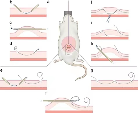
In the constructed mouse excisional wound splint model, the splint ring adheres tightly to the periphery of the wound, preventing localized skin crumpling, and therefore the wound heals again through granulation and epithelial tissue self-repair function, a process that also occurs in humans.
Principle
The excisional wound splint model uses a splint ring that adheres tightly to the skin around the wound, preventing wound closure caused by skin contraction and thus allowing wound healing through granulation and re-epithelialization, a similar process that can be achieved in humans and therefore mirrors human wound healing.
The inner edge of the splint matches the edge of the wound exactly, so the area of the splint hole represents the original wound size, the wound can be photographed at different points in time, and the percentage of wound closure is easy to calculate.
Technical variables caused by skin contraction and wound material are minimized in this model, so uniform wound closure data can be generated for simulating human wound healing.
Appliance
Wound healing, skin regeneration, stem cell and tissue transplantation and immune rejection.
Operation method
Mouse humanoid excisional wound splinting model
Principle
The excisional wound splint model uses a splint ring that adheres tightly to the skin around the wound, preventing wound closure caused by skin contraction and thus allowing wound healing through granulation and re-epithelialization, a similar process that can be achieved in humans and therefore mirrors human wound healing. The inner edge of the splint matches the edge of the wound exactly, so the area of the splint hole represents the original wound size, the wound can be photographed at different points in time, and the percentage of wound closure is easy to calculate. Technical variables caused by skin contraction and wound material are minimized in this model, so uniform wound closure data can be generated for simulating human wound healing.
Materials and Instruments
① BALB/c mice (7~8 weeks old) Move (1) Use a 15 mm diameter circular cutter to cut from a 0.5 mm thick silicone sheet, and then use a 6 mm diameter biopsy punch to cut a small hole in the center to form an annular splint. (2) Carefully clean the splint with detergent and rinse with water. (3) Incubate the splints in sodium hypochlorite (20 000 ppm) for 30 min, rinse with sterile water, and incubate in 70% ethanol for at least 30 min, and air-dry the splints in a tissue culture hood with sterile gauze. (4) Mice were anesthetized by intraperitoneal injection of sodium pentobarbital (50 mg/kg). After anesthesia, the mice were depilated by removing the hair from their backs and flanks, and placed on an experimental table in a supine position to fix the limbs. (5) The skin on the back was cleaned with iodophor and alcohol wipes, and the skin was cut with small scissors. (6) A 6-mm full-length circular wound (extending to the meat sac) was opened on the dorsal side of each animal in a location chosen according to the experiment. (7) The wound holder was opened with silicone rings, which were secured around the wound with adhesive and each ring had eight 6-0 nylon sutures (Ethicon). (8) Dressings were changed every other day under anesthesia. (9) Specific shaped wounds were obtained by excising 10 mm rings of tissue around each wound using a 2 mm biopsy punch, and wound size measurements and healing ratio calculations were performed at d 3, 7, 10, and 14. Caveat 1. Because rodents have different wound healing characteristics than humans, they have distinct limitations. In rodents, because the skin is mobile, skin contraction accounts for a large portion of wound closure; whereas in humans, the skin is connected to the subcutaneous tissue, and new tissue is generated to form the wound healing.The contraction of the skin around the wound is also affected by the posture and movement of the animal, as well as by the wound material, thus leading to considerable variation in the wound closure process.3, A model of wound healing that is stable with minimal technical variables and achieves high reproducibility of data is needed. For more product details, please visit Aladdin Scientific website.
② Sodium hypochlorite
③ Ethanol
④ Sterile water
⑤ Sterile gauze
⑥ Sodium pentobarbital (50 mg-kg-1)