
Nanobody Magnetic Beads
Nanobody Magnetic Beads
In life science research and clinical diagnostics, “specific recognition” and “efficient separation” are two fundamental requirements.Although traditional monoclonal antibodies provide target specificity, they often suffer from limitations such as large molecular weight (~150 kDa), high steric hindrance, and contamination from light and heavy chains. These drawbacks reduce target capture efficiency in complex biological samples.The emergence of nanobody magnetic beads combines the molecular advantages of nanobodies with the separation capabilities of magnetic materials, significantly reducing the limitations of traditional methods and establishing itself as an essential tool in modern biological separation and detection.
I. Unique Advantages of Nanobodies
Nanobodies originate from camelids and are only about one-tenth the molecular weight of conventional IgG. They have a compact structure, are stable in vitro, and retain complete antigen-recognition capability. They offer high affinity, easy engineering, and ease of expression, and can be produced at scale by recombinant methods with high consistency and stable quality.
Leveraging their high affinity, ease of engineering, and in-vitro stability, nanobodies can be orientedly conjugated to magnetic nanoparticles to construct “nanobody magnetic beads.” This tool combines highly specific recognition with rapid magnetic separation: it enables efficient enrichment and elution of targets from complex samples, and—thanks to the very small size and single-domain structure of nanobodies—provides low steric hindrance and facilitates site-specific labeling and multivalency. It is suitable for pathogen detection, exosome/CTC capture, protein purification, and rapid point-of-care testing, while reducing batch-to-batch variation and improving consistency and reproducibility.
II. Principle and Structure of Nanobody Magnetic Beads
1. Basic structure
Nanobody magnetic beads usually consist of three parts:
- Magnetic core: typically Fe₃O₄ (magnetite) nanoparticles with good magnetic responsiveness;
- Surface coating: often a silica (SiO₂)/silica gel layer, polymer, or carboxyl/amino functional layer to improve stability and enable conjugation;
- Nanobody modification layer: specific nanobodies are covalently coupled to the bead surface via chemical conjugation (e.g., EDC/NHS activation, click chemistry), enabling efficient target recognition.
2. Working principle
When nanobody magnetic beads are mixed with a sample, the surface-modified nanobodies bind specifically to target antigens or proteins. An external magnetic field is then used to quickly separate the target complex from the mixture, achieving highly selective capture and purification. This technology combines the high specificity of immune recognition with the high efficiency of magnetic separation.
III. Product Series and Application Scenarios
Product Name | Catalog No. | Main Applications |
Anti-HA Nanobody Magnetic Beads | IP, Co-IP, ChIP, RIP | |
Anti-mCherry Nanobody Magnetic Beads | IP, Co-IP, ChIP, RIP | |
Anti-Flag Nanobody Magnetic Beads | IP, Co-IP, ChIP, RIP | |
Anti-V5 Nanobody Magnetic Beads | IP, Co-IP, ChIP, RIP | |
Anti-GFP Nanobody Magnetic Beads | IP, Co-IP, ChIP, RIP |
Typical application scenarios include:
- Immunoprecipitation (IP/Co-IP): Efficient capture of target proteins and their interacting complexes.
- Chromatin/RNA Immunoprecipitation (ChIP/RIP): Study of DNA/RNA–protein interactions.
- Cell Sorting and Capture: Isolation of circulating tumor cells (CTCs), exosomes, and other rare cell types.
- Rapid On-site Diagnostics: Detection of pathogens and screening of biomarkers.
- Recombinant Protein Purification: High-purity isolation of various tagged proteins.
IV. Aladdin Core Product Advantages
1.Nanobody format, no light/heavy chain contamination. Whether using non-denaturing or denaturing elution, IP complexes will not show contamination from antibody light or heavy chains.
2.High affinity. Low-nM affinity readily handles low-copy genes or hard-to-transfect cell lines.
3.High binding capacity. With oriented coupling, ~15 μg of recombinant protein can be bound per 10 μL of nanobody beads.
4.High specificity. Tested across >10 blank cell lines, the beads show minimal non-specific adsorption.
5.Excellent compatibility. Tags are recognized whether fused at the N- or C-terminus of the bait protein.
6.Good stability. Passed 40 °C stress testing, tolerating temperature excursions in logistics or accidental bench exposure after experiments.
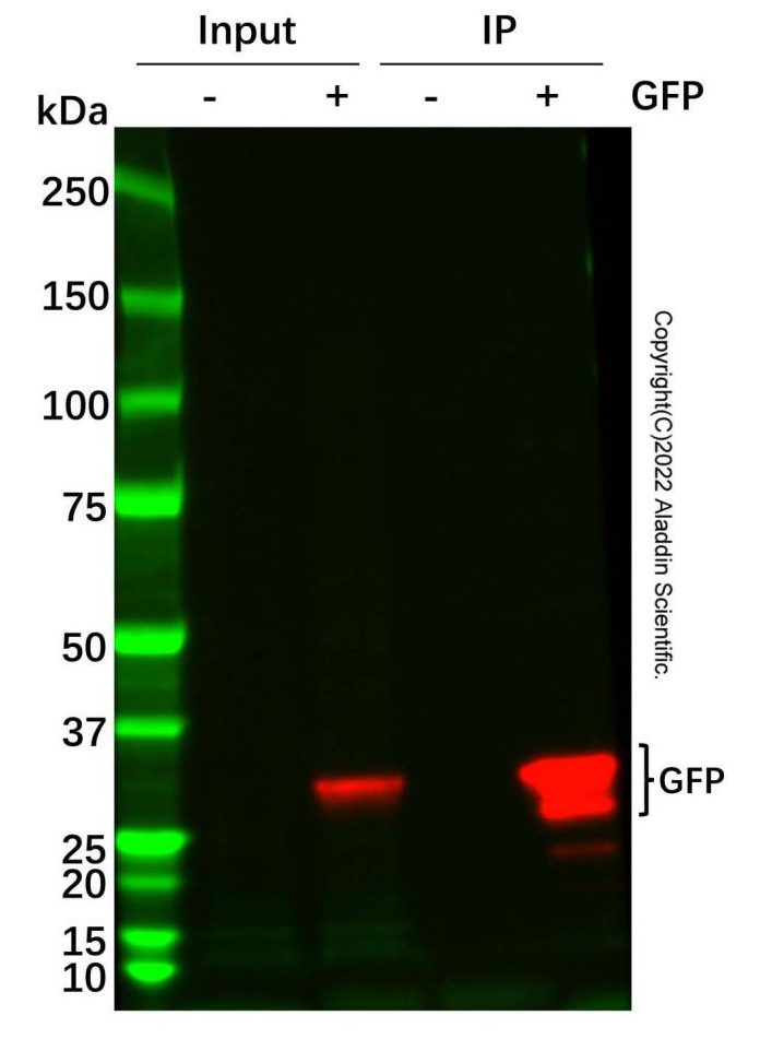
V. Experimental Verification Example: Anti-GFP Nanobody Magnetic Beads (Catalog No.: G1373495)
Western Blot: Anti-GFP Nanobody Magnetic Beads (G1373495)
| All lanes: GFP/EGFP Mouse Antibody (Ab186981) at 1/5000 dilution Lane1: HEK-293T transfected with an empty vector, whole cell lysate Lane2: HEK-293T transfected with an empty vector containing an EGFP-myc-tag, whole cell lysate Lane3: Anti-GFP Nanobody Magnetic Beads (G1373495) IP in HEK-293T transfected with an empty vector, whole cell lysate Lane4: Anti-GFP Nanobody Magnetic Beads (G1373495) IP in HEK-293T transfected with an empty vector containing an EGFP-myc-tag, whole cell lysate Secondary: Goat Anti-Mouse IgG H&L (HRP) (Ab179001) at 1/20000 dilution Predicted band size: 27 kDa Observed band size: 27, 29 kDa Exposure time: 30.0 s |
Nanobody magnetic beads, with their unique molecular recognition capability and convenient magnetic separation, have become an important supporting platform for modern bio-detection and separation technologies. In the future, as nanobody engineering and magnetic materials innovation converge, their applications in disease diagnosis, drug development, and life-science research will become even broader and deeper.
Aladdin: https://www.aladdinsci.com/