
Blue-White Screening of Bacterial Colonies Utilizing X-Gal and IPTG Plates
Blue-White Screening of Bacterial Colonies Utilizing X-Gal and IPTG Plates
Introduction
Blue-white screening of bacterial colonies is a popular and effective molecular biology tool often used to detect recombinant bacteria in cloning experiments. Central to this technique is the enzymatic activity of β-galactosidase, a tetrameric enzyme encoded by the lacZ α gene in E. coli that metabolizes lactose to form glucose and galactose. Alternatively, β-galactosidase can hydrolyze a different substrate, X-Gal, resulting in 5-bromo-4-chloro-indoxyl, which dimerizes to form a blue pigment.
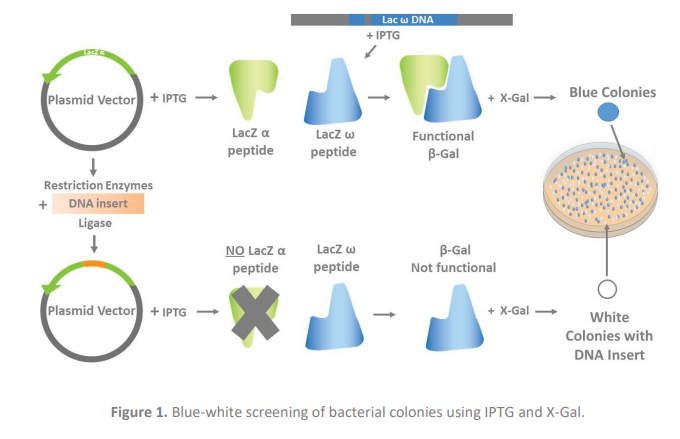
The phenomenon of α-complementation has made β-galactosidase a powerful molecular cloning tool. In α-complementation, the deletion of a specific fragment of the lacZ ω gene in bacteria resulting in an inactive β-galactosidase is resolved by the presence of a plasmid containing the deleted fragment. In cloning, the plasmids routinely used contain a segment of the lacZ α gene, while the E. coli host strain contain a lacZ ω deletion mutation. Thus, during transformation, when bacteria containing the deletion take up the plasmid containing the deleted lacZ α segment, functional β-galactosidase is produced. However, if the plasmid taken up by the bacteria is carrying a piece of DNA (DNA of interest ligated into the plasmid using restriction sites during the cloning process) that disrupts the lacZ α gene segment, recombinant bacteria result. Then, alpha complementation cannot occur, and a functional β-galactosidase does not form.
To perform blue-white screening after transformation, X-Gal is added along with Isopropyl β-D1-thiogalactopyranoside (IPTG), an inducer of lacZ ω gene expression. The blue colonies contain bacteria with functional β-galactosidase, indicating the plasmid taken up during transformation did not contain the DNA of interest. Conversely, the white colonies cannot metabolize X-Gal to produce the blue color, because they do not produce functional β-galactosidase after taking up plasmid carrying the inserted DNA and disrupting the lacZ α gene. These white colonies contain the recombinant bacteria and should be selected (Figure 1). Here, we describe a protocol to perform effective blue-white colony screening to select the recombinant bacteria carrying your DNA of interest.

Materials
· X-Gal (Aladdin Catalog # X274331)
· Dimethylformamide (DMF) (Aladdin Catalog # D111999 etc.)
· dH2O
· Isopropyl β-D-1-thiogalactopyranoside, IPTG (Aladdin Catalog # I274316 I104812 )
· Screening antibiotic of choice
· Agar media (optional)
· Plates
Method
Preparation of X-Gal and IPTG. X-Gal and IPTG can be incorporated into agar media before pouring into plates or added onto pre-made plates.
1. Prepare 20 mg/ml X-Gal in DMF
Note: This stock solution should be stored in a polypropylene or glass tube, protected from
light, at -20°C. This solution is stable for 6-12 months. Aliquots (1 ml) should be made to
prevent degradation due to handling.
Note: For reduced DMF toxicity in media, increase the concentration to 100 mg/ml X-Gal in DMF.
Note: The higher concentration solution is only stable for 1 week at -20°C.
2. Prepare 100mM IPTG solution in dH2O or dilute from a 1M IPTG solution.
This solution is stable for 1 year. Aliquots (1 ml) should be made to prevent degradation due to handling.
Screening on agar media containing IPTG and X-Gal (recommended)
1. Autoclave the growth media agar, then cool to 50°C.
2. Add 10 µl of 20 mg/ml X-Gal solution per 1 ml of media or 2 µl of 100 mg/ml X-Gal solution per 1 ml of media.
Note: recommends using a higher concentration of X-Gal than most protocols as the higher concentration increases blue color intensity and in turn decreases the number of ambiguous colonies needing rescreening. Higher X-Gal concentration also reduces blue color development time and refrigeration time.
3. Add 10 µl IPTG (100mM) per 1 ml of media for a final concentration of 1mM.
4. Add the screening antibiotic.
5. Pour plates and allow them to cool to room temperature before use. This usually takes at least 30 minutes.
6. Spread transformed competent cells as desired.
Note: Blue-white selection plates are generally stable for only 1 week if stored at 4°C in clear sleeves, but may be stored in the dark, or in dark sleeves, at 4°C for up to 1 month.
Screening on pre-made agar plates lacking IPTG and X-Gal
1. Pour autoclaved growth media containing screening antibiotic on media plates and dry in a laminar flow hood.
2. Add 40 µl 100mM IPTG and 120 µl X-Gal (20 mg/ml) to the surface of each plate and spread over the entire surface. Note: The plate edges are difficult to spread evenly and may give false positives. We advise picking colonies in the middle of the plate, if possible, for best results.
3. Dry X-Gal/IPTG-coated media in a laminar flow hood for approximately 30 minutes before use.
4. Spread transformed competent cells and incubate inverted at either 37°C until blue colonies form (usually ~24 hours).
References
Brandt, M. E., Gabrik, A. H. and Vickery, L. E. (1991). A vector for directional cloning and
expression of polymerase chain reaction products in Escherichia coli. Gene, 97(1), 113-117.
Doi:10.1016/0378-1119(91)90017-6.
Juers, D. H., Matthews, B. W. and Huber, R. E. (2012). LacZ β-galactosidase: Structure and
function of an enzyme of historical and molecular biological importance. Protein
Science, 21(12), 1792-1807. Doi:10.1002/pro.2165.
Ullmann, A., Jacob, F. and Monod, J. (1967). Characterization by in vitro Complementation of a Peptide corresponding to an Operator-proximal Segment of the β-Galactosidase Structural
Gene of Escherichia coli. Selected Papers in Molecular Biology by Jacques Monod, 24(2):
339-43. Doi:10.1016/b978-0-12-460482-7.50058-0.
Aladdinsci: https://www.aladdinsci.com