Calcein AM solution - BioReagent, for microscopy, Biological Stain, Suitable for Immunofluorescence(IF), for fluorescence analysis, for cell culture, sterile-filtered, 2 mM in DMSO , CAS No.148504-34-1

CAS: 148504-34-1 Cat. No.: C1456514 Formula: C46H46N2O23 Molecular Weight: 994.87 EC Number: 200-664-3
AVAILABLE TO ORDER
GRADE & PURITY Sterile-filtered ? Sterile-filtered — passed through a 0.2µm filter to remove microorganisms. Use as a ready aseptic solution for culture and sensitive applications. Suitable for Immunofluorescence(IF) ? IF grade — validated for immunofluorescence with low background staining. Use to localize targets in cells/tissue by fluorescence microscopy. BioReagent ? BioReagent grade — tested suitable for life-science and molecular-biology use. Use for cell culture, assays, and biochemical work needing biological compatibility. Biological Stain ? Biological stain grade — dyes characterized for staining cells and tissues. Use in histology and microscopy where staining consistency matters. for Fluorescence analysis ? Fluorescence-analysis grade — very low fluorescent impurities for clean spectra. Use in fluorescence assays where background interferes. for Microscopy ? Microscopy grade — reagents/stains suited to sample prep and imaging. Use in microscopy where clarity and low background are needed. for Cell culture ? Cell-culture grade — low endotoxin and contaminants to support viable cell growth. Use in mammalian/other cell culture media and supplements. 2 mM in DMSO
Synonyms
CAL-AM, CAL-AM solution, Cellstain-Calcein-AM​, Cellstain-Calcein-AM solution, ​CellTrace calcein green AM​, CellTrace calcein green AM solution ​, acetoxymethyl 2-[[2-(acetoxymethoxy)-2-oxo-ethyl]-[[3',6'-diacetoxy-7'-[[bis[2-(acetoxymethoxy)-2-oxo-ethyl
Storage
Protected from light,Store at -20°C,Avoid repeated freezing and thawing
Shipped In
Ice chest + Ice pads
Application
Cell Viability Assay, Flow Cytometry, for microscopy, IF/ICC
★
Size
USA
Germany (EU)*
Price
Qty
100μl
C1456514-100μl
1 In stock
—
$99.90
500μl
C1456514-500μl
US Made to order · 2–4 wks ·
—
$299.90
Enter a quantity for the sizes you want to add.
🧪
Why this grade

BioReagent, for microscopy, Biological Stain, Suitable for Immunofluorescence(IF), for fluorescence analysis, for cell culture, sterile-filtered, 2 mM in DMSO Biological Stain,BioReagent,for Cell culture,for Fluorescence analysis,for Microscopy,Sterile-filtered,Suitable for Immunofluorescence(IF) for sensitive chromatographic and analytical workflows requiring minimal baseline interference.

🌡

Storage & shipping

Protected from light,Store at -20°C,Avoid repeated freezing and thawing Ships Ice chest + Ice pads Check lot-specific COA for exact specifications.

📋

Quality documents

SDS, COA, datasheet, and spec sheet available for download. Lot-specific COA accessible via lot number lookup.

📚

Literature proof

Cited in 129 peer-reviewed publications across chromatography, organic synthesis, and cross-coupling reactions.

Overview

  Calcein-AM is a cell-permeant green fluorescent probe commonly used for determining eukaryotic cell viability or the Mitochondrial Permeability Transition Pore (MPTP). In recent years, Calcein-AM has been widely applied in cell viability analysis experiments.

  Calcein-AM is derived from Calcein by the addition of acetoxymethyl ester (AM) groups, which enhance its hydrophobicity, thereby allowing it to easily penetrate live cell membranes. Calcein-AM itself is non-fluorescent. Once inside the cell, it is hydrolyzed by endogenous esterases to generate calcein, a strongly negatively charged, polar molecule that cannot permeate the cell membrane and is therefore retained within the cell. Calcein emits intense green fluorescence (Ex/Em=494/517 nm). Compared to other similar probes, Calcein-AM is one of the most ideal fluorescent probes for staining live cells due to its very low cytotoxicity—it hardly affects cellular functions such as cell proliferation or lymphocyte chemotaxis—and its low sensitivity to pH changes.

  Since dead cells lack esterase activity, Calcein-AM is used specifically for viability testing and short-term labeling of live cells. The red fluorescent nucleic acid dye Propidium Iodide (PI) can only stain dead cells with compromised membrane integrity, as it cannot penetrate the membranes of live cells. Therefore, Calcein-AM is often used in combination with Propidium Iodide (PI) for simultaneous dual-fluorescence staining of live and dead cells.

Procedure

1. Preparation of Working Solution

  Dilute this product 1:1000 in an appropriate buffer, such as serum-free culture medium, HBSS, or PBS, to prepare the staining working solution.

Note: The optimal final concentration of Calcein-AM is suggested to be adjusted appropriately based on cell type and specific experimental conditions.

2.  Staining Procedure

a. For adherent cells: 

(a) Seed cells in a culture dish, multi-well plate, or on glass coverslips. Treat the cells as required by the experimental design. Gently aspirate the culture medium from the wells and wash with PBS for approximately 10 seconds. Aspirate the PBS.

(b) Add an appropriate volume of Calcein-AM working solution. Gently swirl to ensure even coverage of all cells.

Note: For adherent cells cultured in a 6-well plate with a confluence exceeding 80%, it is recommended to add the staining working solution at a volume of 1 mL per well. This volume can be optimized based on the specific experimental system.

(c) Incubate cells at 37°C for 10-45 minutes. The optimal incubation time varies for different cell types. Consider starting with 10 minutes and optimize for ideal results based on specific experimental conditions.

(d) Aspirate the staining solution and wash the cells 2-3 times with PBS. Then, add serum-free culture medium before observing under a fluorescence microscope. Observe green fluorescence at Ex/Em=494/517 nm. Alternatively, proceed with analysis by flow cytometry or measure fluorescence intensity using a fluorescence microplate reader after staining is complete.

b. For suspension cells: 

(a) Treat cells as required by the experimental design and perform a cell count. Take an appropriate number of cells and centrifuge at 500 x g for 5 minutes at room temperature. Gently aspirate the medium, resuspend the pellet in an adequate volume of PBS, and centrifuge again at 500 x g for 5 minutes at room temperature to remove the PBS.

(b) Add an appropriate volume of Calcein-AM working solution to resuspend the cells at a density of approximately 1x10^6 cells/mL.

(c) Incubate cells at 37°C for 10-45 minutes. The optimal incubation time varies for different cell types. Consider starting with 10 minutes and optimize for ideal results based on specific experimental conditions.

(d) Place a drop of the cell suspension directly onto a glass slide, cover with a coverslip, and observe under a microscope. Observe green fluorescence at Ex/Em=494/517 nm. Alternatively, proceed with analysis by flow cytometry or measure fluorescence intensity using a fluorescence microplate reader after staining is complete.

Note: If background interference is significant, centrifuge to remove the staining solution and wash 1-2 times with PBS before microscopic observation.

Precautions

1.         For your safety and health, wear a lab coat and disposable gloves.

2.         Fluorescent dyes are susceptible to quenching. It is recommended to complete detection on the same day after staining.

Specifications

Synonyms
CAL-AM, CAL-AM solution, Cellstain-Calcein-AM​, Cellstain-Calcein-AM solution, ​CellTrace calcein green AM​, CellTrace calcein green AM solution ​, acetoxymethyl 2-[[2-(acetoxymethoxy)-2-oxo-ethyl]-[[3',6'-diacetoxy-7'-[[bis[2-(acetoxymethoxy)-2-oxo-ethyl
Specifications & Purity
BioReagent, for microscopy, Biological Stain, Suitable for Immunofluorescence(IF), for fluorescence analysis, for cell culture, sterile-filtered, 2 mM in DMSO
Stability And Storage
store at -20°C long term (12 months). Store in the dark.
Storage
Protected from light,Store at -20°C,Avoid repeated freezing and thawing
Shipped In
Ice chest + Ice pads
This product requires cold chain shipping. Ground and other economy services are not available.
Grade
Biological Stain, BioReagent, for Cell culture, for Fluorescence analysis, for Microscopy, Sterile-filtered, Suitable for Immunofluorescence(IF)
Product Line
Biological Products
Images

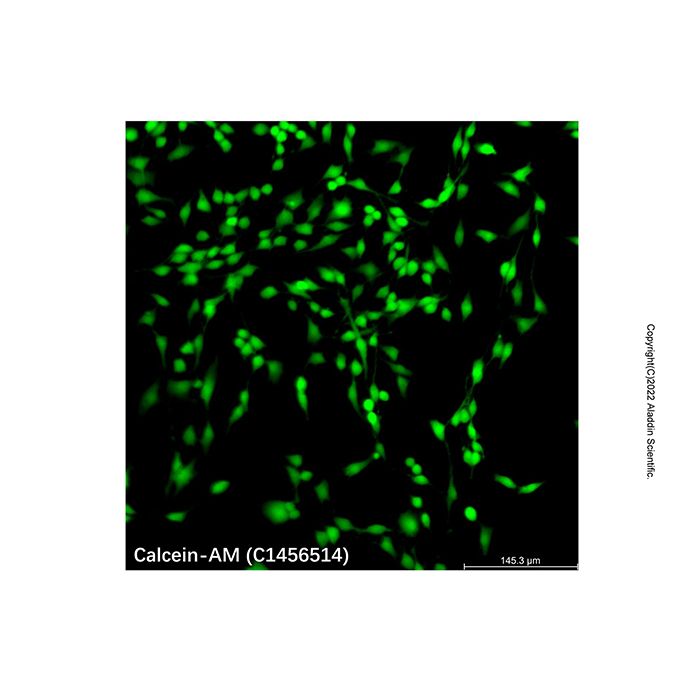
Calcein AM solution (C1456514)
HeLa cells were stained using the Calcein AM solution (C1456514), with the working solution diluted at a ratio of 1/1000 prior to application. Following 10 minutes of staining, the cells were observed under laser scanning confocal microscopy( Ex: 490nm, Em: 515nm ).
Calcein - AM, when entering the cells, is hydrolyzed by intracellular endogenous esterases into Calcein. Calcein is a polar molecule with a strong negative charge, unable to pass through the intact cell membrane, so it stays within the cells and emits intense green fluorescence.
In the image, the bright green fluorescent regions correspond to metabolically active live cells. These cells have intact morphology, with clearly distinguishable cell bodies and protrusions, indicating that the cellular structure and viability of HeLa cells are well preserved under the experimental conditions.

Names and Identifiers
Isomeric SMILES
CC(=O)OCOC(=O)CN(CC1=CC2=C(C=C1OC(=O)C)OC3=C(C24C5=CC=CC=C5C(=O)O4)C=C(C(=C3)OC(=O)C)CN(CC(=O)OCOC(=O)C)CC(=O)OCOC(=O)C)CC(=O)OCOC(=O)C
Molecular Weight
994.87
Reaxy-Rn
10757520
Reaxys-RN_link_address
https://www.reaxys.com/reaxys/secured/hopinto.do?context=S&query=IDE.XRN=10757520&ln=

Documentation

📋 Safety Data Sheet (SDS)

Comprehensive hazard, handling, storage, and regulatory compliance document.

<<<<<<< HEAD Download SDS → ======= Download SDS → >>>>>>> release248-p3

✅ Certificate of Analysis (COA)

Lot-specific quality data. Enter your lot number to retrieve the exact COA.

Look up COA →

📊 Datasheet

Quick-reference summary of product specifications and applications.

View datasheet →

🔬 Specification Sheet

Full quality attributes and acceptance criteria for this grade.

View spec sheet →

Advanced Data

Certificates (CoA,COO,BSE/TSE and Analysis Chart)
C of A & Other Certificates(BSE/TSE, COO):
Analytical Chart:

Find and download the COA for your product by matching the lot number on the packaging.

7 results found

Lot NumberCertificate TypeDateItem
ZJ26F0939280ZJ26F0939280Certificate of AnalysisSep 17, 2026 C1456514
ZJ26F0939279ZJ26F0939279Certificate of AnalysisSep 17, 2026 C1456514
ZJ25F1029081ZJ25F1029081Certificate of AnalysisAug 08, 2026 C1456514
ZJ26F0636524ZJ26F0636524Certificate of AnalysisJun 16, 2026 C1456514
ZJ26F0636523ZJ26F0636523Certificate of AnalysisJun 16, 2026 C1456514
ZJ26F0434465ZJ26F0434465Certificate of AnalysisApr 21, 2026 C1456514
ZJ26F0434464ZJ26F0434464Certificate of AnalysisApr 21, 2026 C1456514
Chemical and Physical Properties
SensitivityLight-sensitive
Documents & Articles
Sterile-Filtered
Suitable for Immunofluorescence(IF)
Biological Stain
Choosing Reagents ‘for Fluorescence Analysis’: A Practical Guide
For Cell Culture
Cell Live/Dead Staining and Apoptosis Detection Techniques
Principles and Methodological Framework for Nucleic Acid Staining and Organelle Labeling
Fluorescent Probes: A Complete Beginner’s Guide — Definitions & Emission Mechanisms, Signal Readouts, Classification Framework, Selection Roadmap, and Product Tables (Tables 1–4)
Comparative Guide and Selection Framework for Common Fluorophores
Oxidative Stress and Experimental Interpretation Framework for ROS, JC-1, MPTP, and Calcium Indicators in Mitochondrial Function Assessment
Application Comparison of the Neutral Red Assay and the MTT/CCK-8 Assays in Cytotoxicity Testing
Comparison of Common Mammalian Cell Antibiotic Selection Systems: Puromycin, G418, Hygromycin B, and Blasticidin
Construction and Evaluation Strategy of a Gentamicin-Induced Cell Injury Model
Summary of Cell Staining Methods
Citations of This Product
Solution Calculators
Reviews

Customer Reviews

Frequently Asked Questions

What does sterile-filtered mean?
sterile-filtered indicates that the material has been processed and tested for specialty industrial processing. Specifications focus on the impurities that affect performance in that application — for example, metal content for electronics, particle size for batteries, refractive index for optics, or moisture content for moisture-sensitive processes.
What does Suitable for Immunofluorescence(IF) mean?
Suitable for Immunofluorescence(IF) indicates the product has been validated for use in immunoassays such as ELISA, IF, IHC and flow cytometry. Validation may include positive/negative control samples, signal-to-noise testing, and protocol-specific QC. Lot-specific test data is published on the Certificate of Analysis where available.
How should this product be stored?
Store at ?20 °C, protected from light. Avoid repeated freeze–thaw cycles. Aliquot into single-use volumes before freezing.
How is this product shipped?
This product ships in an insulated container with ice pads. Unpack on arrival and transfer it to the storage condition stated above.
What are the CAS number, molecular formula and molecular weight?
The CAS Number is 148504-34-1, the molecular formula is C46H46N2O23, and the molecular weight is 994.87 g/mol.
What documentation is provided?
Available product documentation, including Certificates of Analysis (COA), Safety Data Sheets (SDS), and specification sheets, is shown in the product document area. Document availability and access follow the current site policy.

Shall we send you a message when we have discounts available?

Remind me later

Thank you! Please check your email inbox to confirm.

Oops! Notifications are disabled.