
Observations on the basic structure and posterior contents of plant cells
Observations on the basic structure and posterior contents of plant cells
Cells are the basic units that make up living organisms. Compared with animal cells, plant cells have their own special cell components, such as cell wall and plasmodesmata. Normally, from the outside to the inside, the cell wall can be divided into three layers: the intercellular layer, primary wall and secondary wall. The main component of the intercellular layer is pectin, which is secreted outward by two neighboring protoplasts in the early stage. Pectin can be stained red by nail red. The primary wall is mainly composed of cellulose, hemicellulose and pectin. Primary walls containing cellulose can be stained blue-purple with zinc chloride-iodine solution. The primary wall can be shown by this method.
The secondary wall is the wall material, often containing lignin, that is secreted to form on the inside of the primary wall after the protoplast stops growing. Lignin can be stained red by resorcinol, so this method can be used to show the presence of secondary walls. Plastids are organelles that are unique to plant cells and can differentiate to form chloroplasts, colored bodies, and so on. Both chloroplasts and colored bodies are large organelles and can be observed under a light microscope. Chloroplasts are found mainly in the chloroplasts and contain the pigments chlorophyll a and chlorophyll b. Because these two pigments are weak absorbers of green light, chloroplasts reflect a lot of green light and appear green. Colored bodies lack chlorophyll and contain carotenoids, etc.
According to the content and composition of these pigments, the colored bodies appear red, yellow and other colors, such as the red color of red pepper fruits is due to the presence of a large number of colored bodies. The after-containing material is the cell in the process of growth and differentiation, as well as mature due to metabolic activities produced by the storage material or waste. Some of them are present in the vesicles, and some of them are present in the organelles.
Operation method
Observations on the basic structure and posterior contents of plant cells
Materials and Instruments
Materials: Move The basic process of observing the basic structure and posterior contents of plant cells can be divided into the following steps: 1. Cellulose Cellulose is the predominant component of the cell wall and is a long chain of chemicals formed by the dehydration and condensation of many glucose molecules. Take a clean slide and place a drop of water in its center with a dropper. Take a fresh onion leaf and tear a small piece of epidermis (5 mm) from the inside of the onion bulb with a razor blade or shackle and quickly place it in the drop of water in the center of the slide. If curling occurs, it should be carefully unrolled with a dissecting needle and covered with a coverslip. Note that when covering the coverslip, one side of the coverslip should be clamped with a spatula so that the other side is in contact with the edge of the water droplet, and slowly lowered until it is flat (the air is squeezed out to avoid air bubbles). The cell wall characteristics of the inner epidermis of the onion bulb in its natural state were observed under a microscope. Then, the staining treatment of iodine-potassium iodide solution was carried out on the slide: 1% of iodine-potassium iodide solution was taken and added to the material, and then 1 drop of 66.5% sulfuric acid was added, and cellulose in the cell wall showed blue color by the action of iodine and sulfuric acid. The cellulose-containing primary wall was observed to be blue-violet under the microscope. The more cellulose in the cell wall, the more obvious the blue color; in the secondary wall, the fibers have been covered by lignin, etc., and do not change to blue. A drop of I2-ZnCl2 reagent can also be applied to plant material, and cellulose-containing cell walls appear bluish-purple. 2. Pectin Pectin is the main substance that constitutes the intercellular matrix of higher plants, and is the main chemical component of the intercellular layer and the primary wall of dicotyledonous plants, with a small amount in the cell walls of monocotyledonous plants. The epidermis of onion bulbs was stained with nail red solution using the method described above (time 30 min). Under the microscope, the intercellular layer between the two primary walls was seen to be red in color. 3. Lignin Lignin is a polymer of aromatic compounds. The thin slices of cut lignocellular stems were placed on slides and then stained with hydrochloric acid resorcinol solution. The slices were dipped in hydrochloric acid dropwise for 3~5mim and then 5%~10% of resorcinol ethanol solution, and the red lignified cell walls were visible under the microscope. Depending on the depth of color, it can show the degree of lignification in the cell wall.
Onion bulbs, red pepper fruits, tobacco leaves, duckweed leaves, potato tubers and woody stems, young roots or young stems of plants, peanut seeds, soaked wheat and maize seeds, grate hemp seeds, persimmon endosperm permanently sliced, pine stems sliced in three sections.
Apparatus:
① Microscope
① Microscope ② Slide
① microscope ② slide ③ coverslip
① microscope ② slide ③ coverslip ④ tweezers
① Microscope ② Slide ③ Coverslip ④ Tweezers ⑤ Dissecting needle
⑥ Absorbent paper
⑦ Gauze
Reagents:
① Distilled water
② Zinc chloride and iodine solution
① Distilled water ② Zinc chloride and iodine solution ③ 66.5% aqueous nail red sulfate solution
④ Resorcinol hydrochloride solution
⑤ Iodine-potassium iodide solution
⑥ Hydrochloric acid
⑦ Sulfuric acid
⑧ Sudan III
1. chloroplast
Tear a piece of tobacco leaf epidermis under a dissecting microscope and place it on a slide for observation. Under the microscope, you can see the epidermis with some green cells, these are torn epidermis brought down when the chloroplasts. Observe carefully the morphology of chloroplasts in the chloroplasts.
2. colored bodies
Cut a thin slice of red pepper with a razor blade. Under the microscope, observe the orange colored particles.
3. White bodies
Tear the epidermis of a duckweed leaf and make a clinical slide for observation. First, find the epidermal cells and guard cells and vice-guard cells under low magnification, and then switch to high magnification, in the vice-guard cells around the nucleus there are some colorless and transparent orb-like particles that is white body.
(iii) Observation of after-containersPosterior content is mainly storage material, mainly starch granules, lipids, paste powder granules.
1. starch grain
Starch is the most common post-container in plant cells, mainly stored in the form of starch granules. Take a small piece of potato tuber, use the blade to cut off the surface oxidized layer, scrape a little juice, made of temporary water mounted film, placed in a low magnification microscope observation, can see a lot of different sizes of particles, that is, starch granules. Select the granules are not dense and do not overlap each other, with high magnification observation. Since the starch granules were unstained, it was necessary to adjust the aperture size and fine focus to observe clearly. The oval-shaped amyloplasts have alternating dark and light concentric whorls arranged eccentrically around a core (umbilicus) (Figure 11-1A). The majority of amyloplasts in the field of view are single grains with a single umbilicus, and there are a small number of complex and semicomplex grains with two or more umbilicus points. The central portion of the semicomplex grains had each umbilicus with its own whorl and a common concentric circle around the periphery; the umbilicus of the complex grains had only its own whorl and no common concentric circle. When a small amount of iodine-potassium iodide solution is added dropwise from one side of the coverslip and absorbed from the other side, the iodine-potassium iodide solution gradually enters under the coverslip, and the starch grains are stained blue or purple because starch shows blue or purple coloration when exposed to iodine (Figure 11-1B).

Compare the starch grains of endosperm cells of wheat, corn, and other plants. Take wheat and corn kernels that have been soaked, cut some of the endosperm cells with bare hands, pick the thinnest piece, place it on a slide, and make a clinical slide in the same way as above to compare how they differ from potato starch grains in shape, size, and structure.
2. Paste grain
Paste granules are the storage places for proteins in plant cells, and are often found in the cells in an amorphous or crystalline state. Take wheat or corn seed slices, in the endosperm of the outermost layer to find paste powder layer, paste powder layer cells nearly square, arranged more neatly, the cells have many stained or colorless small round particles that are paste powder grain. There is also a class of large composite dextrin particles, take the fungus hemp seeds, peel off the patterned seed coat, with the fat endosperm to do freehand section, a thinner piece on the slide, the first drop of 95% ethanol in order to dissolve the fat, and then drops of iodine-potassium iodide solution staining, sealing the film, observed with a low-power objective, can be seen in the thin-walled cells are full of oval oval large composite dextrin particles are stained yellow. High-power objective to observe the structure of a paste grain: the outer protein membrane, the inner package of 1 to several polygonal crystals, i.e., protein molecules, stained dark yellow, and a colorless, unstained spherical crystals, which is not a protein molecule, but inorganic phosphate compounds and calcium, magnesium and other salts combined. Alternatively, it can be detected by the mercuric chloride-mesophenol blue method, in which 1 drop of mercuric chloride-mesophenol blue is added to a section, and after 5 min the excess dye on the section is rinsed off with 0.5% acetic acid, and then placed in a petri dish and washed in water for 5 min, and then sealed with glycerol, and the pasty powdery particles in the cells are stained bright blue.
3. Oil droplets
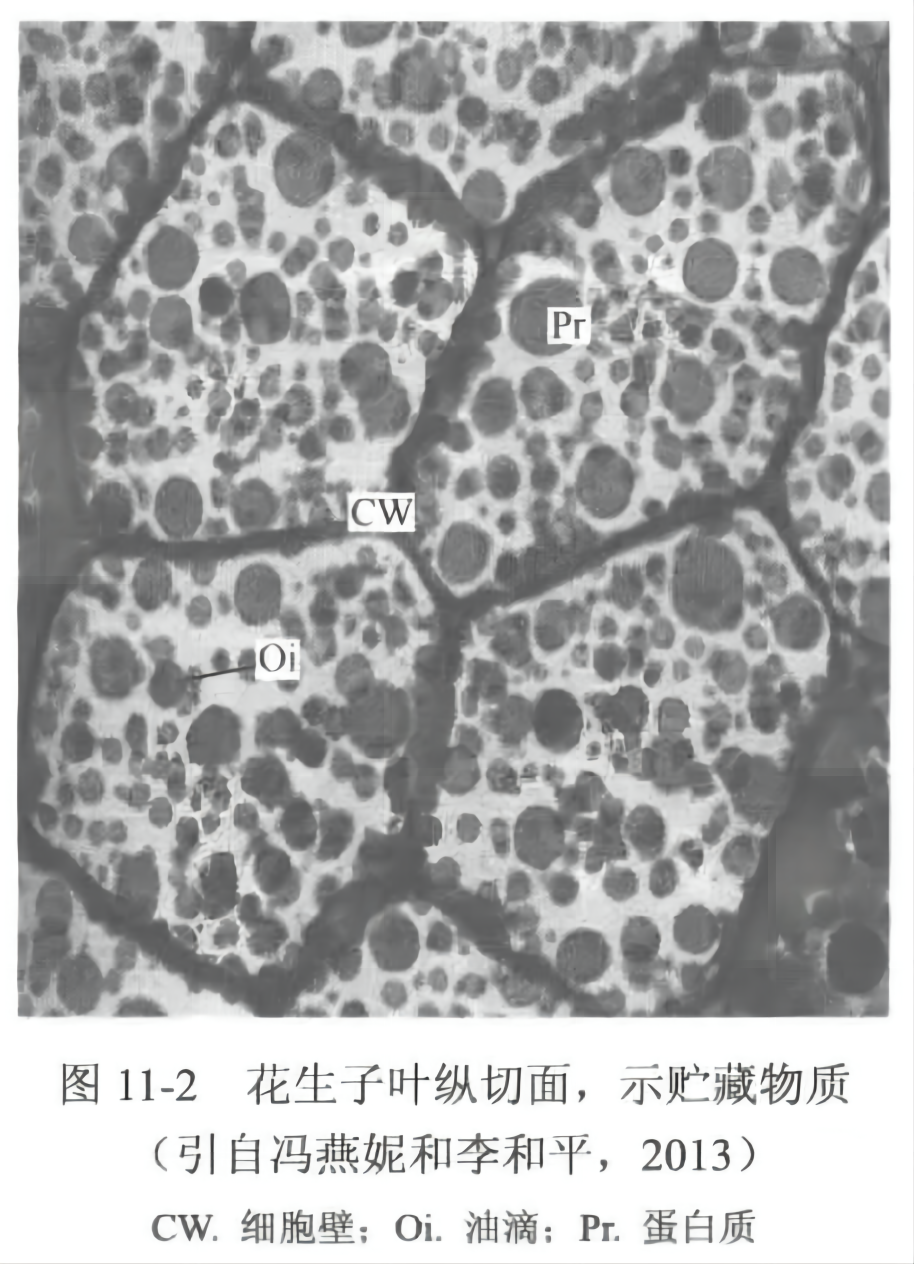
The fat stored in plant cells often exists in the form of oil droplets. Take a peanut seed, peel off the red seed coat, use a razor blade to cut a very thin section, put it on a slide, add drops of Sudan Ⅲ (or Sudan Ⅳ) ethanol solution staining for more than 15 min, make a fashion slide, put it under the microscope to observe, you can see that there are rounded oil droplets inside the peanut cotyledon cells are stained with orange-red color (Fig. 11-2), and the embolus and the cuticle are stained similarly. The fat showed purple-red color in the presence of comfrey reagent and turned black in the presence of tetroxide starvation.
4.3 Comprehensive identification of post-species contents
Take the peanut slices with oil droplets as materials, add iodine-potassium iodide solution on the basis of Sudan Ⅲ staining, absorb the overflowed liquid, observe the stained slices, and at the same time, you can see blue-purple starch granules, light-yellow protein nuclei, and orange-red oil droplets, with this method, you can study the content and distribution of three different storage substances in the cells.
For more product details, please visit Aladdin Scientific website.