Blood-brain barrier permeability assay
Blood-brain barrier permeability assay
Evans blue staining is of very wide use in in vivo experiments in small animals involving the cardiovascular system, nervous system, skeletal muscle system and other diseases in the research method, here we focus on Evans blue staining in the detection of blood-brain barrier permeability in the detection method and application.
Principle
Evans Blue is a commonly used azo dye preparation that has a high affinity for plasma albumin, which is unable to cross the blood-brain barrier under physiological conditions, and Evans Blue bound to plasma albumin is unable to color it. When the blood-brain barrier of the nervous system is destroyed, Evans blue can enter the nervous system and color it. Evans blue dye has a strong peak at fluorescence wavelengths of 470 and 540 nm, and its levels in tissues are often detected using chemotaxis and colorimetric methods.
Evans blue perfusion staining combined with confocal laser scanning microscopy to observe the fluorescence intensity of Evans blue dye in brain slices can be used to detect alterations in the morphology of blood vessels in the blood-brain barrier and to quantify the amount of Evans blue dye infiltrated into brain tissue.
Operation method
Evans Blue assay for blood-brain barrier permeability
Principle
Evans Blue is a commonly used azo dye preparation that has a high affinity for plasma albumin, which is unable to cross the blood-brain barrier under physiological conditions, and Evans Blue bound to plasma albumin is unable to color it. When the blood-brain barrier of the nervous system is destroyed, Evans blue can enter the nervous system and color it. Evans blue dye has a strong peak at fluorescence wavelengths of 470 and 540 nm, and its levels in tissues are often detected using chemotaxis and colorimetric methods. Evans blue perfusion staining combined with confocal laser scanning microscopy to observe the fluorescence intensity of Evans blue dye in brain slices can be used to detect alterations in the morphology of blood vessels in the blood-brain barrier and to quantify the amount of Evans blue dye infiltrated into brain tissue.
Materials and Instruments
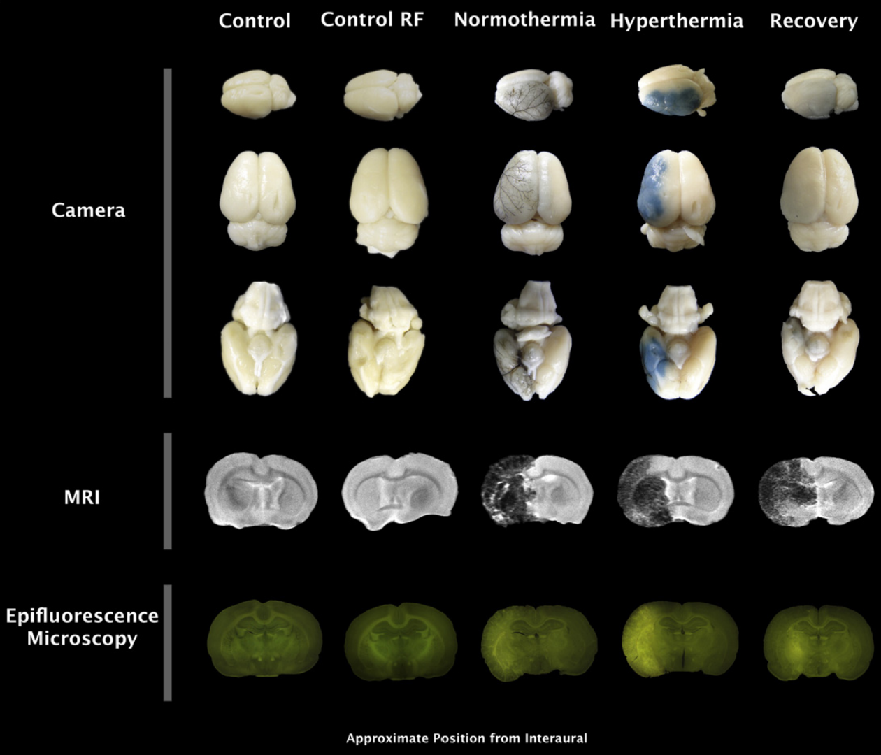
Small animals to be tested (mice, rats, etc.), 2% Evans blue stain, anesthetic, 0.9% sodium chloride solution, heparin, 1 mL syringe, ophthalmic scissors, forceps. Move The steps of Evans blue assay for blood-brain barrier permeability were as follows: 1. Prepare 2% Evans blue dye solution (2 g + 100 mL of double-distilled water) and sodium chloride solution containing 20 U/mL heparin. 2. Anesthetize the mice with an anesthetic (anesthetic is not recommended here; choose the appropriate dose according to the weight of the mice). 3: Inject 2% Evans Blue dye solution (2 mL/kg) into the tail vein or femoral vein. 4: Open the chest cavity of the mouse and slowly inject 0.9 sodium chloride solution (containing heparin) from the apical portion of the heart, and cut open the right atrium until the fluid coming out of the right atrium is clarified, and then stop the perfusion. 5、Separate the brain tissue, if there is obvious coloration, can be directly photographed with a body microscope 6、After taking general photographs, the brain tissue can be cut according to the situation and demand, and used to prepare frozen sections for confocal photographs, or take the supernatant after homogenization at 620 nm for spectrophotometric measurement. J Control Release. 2015 May 28;206:49-57. doi: 10.1016/j.jconrel.2015.02.027. Epub 2015 Feb 25. In the figure, Camera is the photographed image under the body microscope, Epifluorescence Microscopy is the photomicrographs under fluorescence microscopy. Caveat 1. It is better to calculate the dosage of Evans blue dye solution in advance and use it now. 2. For statistical analysis, the value measured by spectrophotometer is the absorbance, if you want to further quantify, you need to prepare the standard in advance and construct a standard curve for quantitative analysis. 3. The fluorescence pictures taken by confocal can be analyzed by imageJ for relative quantitative analysis. Common Problems 1. What if the tail vein injection of Evans Blue Stain is not easy to succeed? If you are not skilled in tail vein injection, you can inject through the femoral vein or jugular vein of the mouse. 2. Does the fact that the brain tissue (with damaged blood-brain barrier) does not stain well (no blue color visible to the naked eye) mean that the experiment has failed? Don't worry, although there is no visible blue color on the brain tissue, it does not necessarily mean that the experiment has failed, you can homogenize the tissue and take the supernatant for further quantitative analysis by absorbance photometer. For more product details, please visit Aladdin Scientific website.