
GENERAL PROTOCOL OF DYE MALEIMIDE ANTIBODY LABELING KIT
GENERAL PROTOCOL OF DYE MALEIMIDE ANTIBODY LABELING KIT
Overview
The BroadPharm Dye Maleimide Antibody Labeling Kits contain sulfonated maleimide dyes which label thiolcontaining proteins, including antibodies. The kit provides optimized reagents for labeling antibodies having 2.5-3 labels per antibody and desalting columns for purifying the labeled molecule. The dye maleimide, a controlled excess component, reacts with thiol groups of the reduced antibody at neutral pH. The purification of the antibody is achieved by size filtration using centrifugal concentrator or SEC column.
The 100 μg kit provides sufficient dye for labeling 50-200 μg of antibody in ~100 μl volume, and the 1 mg kit can label 1-5 mg of antibody in ~ 500 μl volume.

AT A GLANCE
Protocol summary
• Prepare the antibody for labeling.
• Optional: Reduce disulfide bonds with TCEP (~30 minutes).
• Prepare the dye stock solution.
• Perform the labeling reaction (2 hours).
• Purify the conjugated antibody.
• Calculate the degree of labeling.
• Add antibody stabilizers to conjugate.
Note: Upon receipt, store components NHS at -20°C and component B at 40C. Before use, allow all the components to reach room temperature, centrifuge the vials briefly before opening, and immediately prepare the required solutions prior to starting your conjugation. The following SOP is an example for labeling goat anti-mouse IgG antibody.
PREPARATION OF ANTIBODY WORKING SOLUTION
1. Preparation of antibody of antibody working solution: the preferred antibody concentration is 2 mg/ml in supplied buffer component B, or PBS buffer pH 7.2-7.4.
2. Optional: reducing antibody with TCEP
a. Dissolve antibody in 1X PBS at concentration around 2 mg/ml.
Add ~10-fold molar excess of TCEP.
b. Incubate the reaction solution for ~30 minutes. The reduction reaction and subsequent labeling reaction are best carried out in the presence of an inert gas (N2 or Ar) to prevent re-formation of disulfide bonds.
Note:
• TCEP as well as other reducing agents such as DTT cross react with the maleimide moiety and need to be removed (i.e. by dialysis or spin filtration) prior to labeling. Residual of TCEP will lower the labeling efficiency.
• Adjust the antibody concentration to 2mg/ml in reaction buffer if needed. That is, 200 μg antibody in 100μl reaction buffer for 100μg kit and 1mg antibody in 500μl reaction buffer.
• The antibody should be dissolved in supplied buffer component B, or 1X phosphate buffered saline (PBS), pH7.2-7.4. If the antibody is dissolved in glycine buffer, it must be dialyzed against 1X PBS, or use Amicon Ultra-0.5, 10 kDa molecular weight cutoff (MWCO), for desalting (Cat # UFC501008 from Millipore Sigma). These methods, can also be used remove free amines or ammonium salts (such as ammonium sulfate and ammonium acetate) that are widely used for antibody precipitation.
• Impure antibodies or antibodies stabilized with bovine serum albumin (BSA) or gelatin will not be labeled well.
• Labeling efficiency varies with antibody concentration. For optimal labeling efficiency, a final antibody concentration range of 1-10 mg/ml is recommended. In general, the higher the antibody concentration,the better the labeling efficiency of the dye. This protocol is designed to yield optimal degree of labeling with antibody concentration range 1-2 mg/ml.
SAMPLE EXPERIMENTAL PROTOCOL
1. For 100 μg kit, prepare antibody stock at a concentration of 2 mg/ml in supplied buffer component B, or PBS buffer pH 7.2-7.4. Aliquot 100 μl into a vial for use.
For 1 mg kit, scale the antibody content to desire concentration for reaction volume ~500 μl max.
2. Calculate appropriate volume of reactive dye stock solution.
Note: for antibody lgG (150KDa), the recommended mol ratio dye: antibody is 10 to 20.
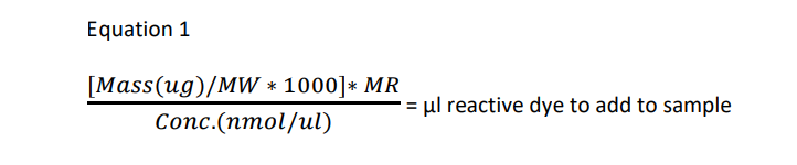
Where:
Mass: antibody mass (μg)
MW: antibody molecular weight
MR: is the dye: antibody ratio from the 10 to 20.
Conc.: the concentration (nmol/μl) of the reactive dye stock solution prepared below.Note: 2 nmol/μl for 100 μg kit; 10 nmol/μl for 1mg kit.
3. Allow a vial of dye maleimide vial to warm up to room temperature, and then add 50 μl anhydrous DMSO to it.Vortex to dissolve the dye. The concentration of this reactive dye stock solution is 2 nmol/μl for 100 μg kit; 10nmol/μl for 1mg kit.
4. Add the appropriate volume of reactive dye solution, based on equation 1, to antibody buffer vial and mix well. Protect the antibody/dye solution from light by wrapping the vial in aluminum foil and incubate the reaction for 1 hour at room temperature with gentle rocking.
5. For most of the bio-application, optimal DOL for each dye is listed here for reference.
|
Note: the DOL is depend on antibody conc, reactive dye/ antibody ratio, and free -SH amount in antibody. Please use the table to optimize labeling condition (MR) and reduction condition (TCEP/DTT amount and incubation time) accordingly if higher DOL is needed.
Buffer Exchange and Excess dye Removal
100 μg kit excess dye removal by spin column:
1. Remove bottom closure of Zeba Spin desalting column, and then loosen but do not remove the cap.
2. Place column in a 1.5-2.0 ml collection tube (microcentrifuge tube). Centrifuge at 1,500 × g for 1 minute to remove storage solution.
3. Place a mark on the side of the column where the compacted resin is slanted upward. Place column in the microcentrifuge with the mark facing outward in all subsequent centrifugation steps.
4. Transfer the column to a new collection tube, uncap to apply labeling reaction sample to the top of the compact resin bed, slowly (volume not to exceed a 130 μl).
5. Centrifuge at 1,500 × g for 2 minutes to collect the sample. Discard the desalting column after use.
6. Store dye-antibody at 4°C for < 1 month. For longer periods, store at -20°C or -80°C, optionally with stabilizers (e.g., 0.1% bovine serum albumin) and 0.02-0.05% sodium azide.
1 mg kit excess dye removal by protein concentrator:
1. Hydrate concentrator membrane ‘filter device’ with 400 to 500 μl of reaction buffer or DI water, and microcentrifuge 14,000 x g, for 3 minutes. Discard, liquid from filter device and collection tube.
2. Spin down by adding the dye-antibody to the concentrator/filter device up to 500 μl. Microcentrifuge at 14,000 x g, 8 minutes, or to minimum volume ~ 50 μl left in the filter device. Discard waste from the collection tube.
3. Desalt by adding reaction buffer to the filter device up to 500 μl total volume; usually ~450 μl of buffer.Microcentrifuge at 14,000 x g, 8 minutes, or to minimum volume ~ 50 μl left in the filter device. Discard waste from the collection tube.
4. Repeat step 3, twice.
5. Collect dye-antibody from the filter device into a microcentrifuge tube.
6. Optional for maximum recovery, add a small volume of buffer component B, determined by the user, to the filter device to rinse out residual antibody, microcentrifuge pulse spin, collect antibody/reaction buffer from filter device, add to the microcentrifuge tube from step 5, mix.
Dye/Protein ratio calculation
To calculate dye to antibody ratio, measure absorption spectrum of the conjugate, or absorption at 280 nm (AAb), and at dye absorption maximum (Adye). A typical absorption spectrum of a dye labeled antibody is shown below. Depending on the dye, wavelength of the maximum may vary.
|

A ᵈʸᵉ : Absorbance of dye
Ɛ ᵃᵇ : Molar extinction coefficient at 280 nm of biomolecule
A ᵃᵇ : Absorbance at 280 nm of the dye-protein conjugates
CF: Correction factor at 280nm accounting for the absorption of the dye at 280 nm
Ɛ ᵈʸᵉ : Molar extinction coefficient of the dye
Storage of Antibody-Dye
The antibody conjugate should be stored in the presence of a carrier protein.
Troubleshooting
|
FAQ:
Q: What is the mechanism of the thiol-maleimide reaction?
A:

Figure 3 shows the general mechanism of the thiol-maleimide reaction. The high specific reactivity of the olefin is due primarily to (a) the ring strain arising from the bond angle distortion and (b) the positioning of the carbonyl groups in the cis-conformation. With highly polar solvents such as water, dimethyl sulfoxide (DMSO), N,N’-dimethylformamide (DMF), or N,N’-dimethylacetamide (DMAC), the thiol-maleimide reaction proceed without a catalyst because of the formation of the active species the thiolate ion.
Q: What is the right pH for this reaction?
A: From pH 6.5 to pH 7.5, the thiol-maleimide reaction is chemoselective for thiols. At pH 7.0, the reaction rate of maleimide with thiol is about 1,000 times faster than the reaction rate of maleimide with amine. However, above pH 7.5, free primary amines react competitively with thiol at the maleimide C=C bond.

Q: Is the thio-succinimide conjugate stable?
A: In aqueous solution, the maleimide ring can be opened by hydrolysis. This susceptibility to hydrolysis increases with increasing pH. Importantly, if the ring-opening reaction occurs before thiolation, the resultant maleic amide is unreactive to thiol. On the other hand, if thiolation has occurred, the ring-opened succinamic acid thioether is stable. Because of the propensity for ring-opening hydrolysis and inactivation, we do not recommend aqueous storage for products containing a maleimide, which is intended for future reactions. If solution storage is required, use a dry, water-miscible, biocompatible solvent such as DMSO, DMF, or DMAC).

Dye maleimide products are easy to use effectively following our recommended instructions on the product information sheets.
The following are some recommended best practices for working with our dye maleimide products.
• Products should be stored at the recommended temperature except when in use. The recommended storage temperature for almost all products that contain maleimide is -20°C.
• When a product containing maleimide is removed from storage, it should be allowed to equilibrate fully to ambient temperature before opening the bottle or vial in which the product is stored.
• Aqueous solutions of maleimide-containing products should be made immediately before use. If the maleimide functional group is to be reacted, the pH should be 6.5 – 7.5, and preferably as low as possible within that range.
• Aqueous buffers and organic solutions of maleimide-containing products should be free of primary and secondary amines and free of thiols. If a base need to be used in a reaction with a maleimide-containing product, we recommend a highly hindered organic base such as 2,6-lutidine (CAS number 108-48-5; EC number 203-587-3).
• Do not store maleimide-containing products in aqueous solutions due to the risk of hydrolysis. Instead, use a dry, biocompatible, water-miscible solvent (g., DMSO, DMF, or DMAC) for the long-term storage of these compounds. These solvents can be dried suitably over 3 Å molecular sieves (8×12 mesh recommended) for 24 – 48 hours at 20 – 25°C. Solubilized maleimide-containing products should be stored at -20°C. We do not have information on the stability of maleimide-containing products stored in solution because we explicitly do not recommend such storage for these products.
• When conjugating a maleimide containing product to a protein, if a stock solution of the maleimidecontaining product in an organic solvent (vide supra) is added to the reaction mixture, no more than 10% of the final reaction volume should be the organic solvent, while the rest of the volume should be water or aqueous buffer (for example, PBS). Some sensitive proteins may require that the amount of organic solvent be much less than 10% of the final reaction volume.
Aladdinsci: https://www.aladdinsci.com