
PCR-Denaturing Gradient Gel Electrophoresis
PCR-Denaturing Gradient Gel Electrophoresis
PCR-denaturing gradient gel electrophoresis is the process of amplifying the target gene through PCR by adding a nucleotide chain of 30-50 GC bases to the 5' end of one of the primers, so that one end of the amplified product contains GC bases, i.e., the GC-clamp. since the GC-clamp is the region with the highest Tm value in the amplified fragment, while the other parts of the amplified fragment have different Tm values due to the different types of bases, and are obviously lower than the highest Tm value. Since the GC-clamp is the region with the highest Tm value in the amplified fragment, the Tm values of other parts are different depending on the types of bases they contain, and are obviously lower than the highest Tm value. When the GC-clamped DNA fragment is electrophoresed in a polyacrylamide gel with a linear gradient of denaturants (urea and formamide), the double-stranded DNA is partially unstranded when it reaches a certain concentration of denaturing region (the denaturing effect of this concentration of denaturant corresponds to the Tm value of the region of the DNA fragment with the lowest unstranding temperature). The partially unstranded DNA molecules become branched and migrate at a significantly slower rate, eventually coming to a virtual standstill. Whenever a base in the DNA fragment is mutated, it leads to a synergistic effect between the bases, which causes a change in the Tm value, and partial unlinking occurs at different concentrations of denaturant, resulting in a near-stop of swimming, and ultimately in the detection of mutations in the genes. At present, there is one main method for PCR-denaturing gradient gel electrophoresis: PCR-denaturing gradient gel electrophoresis.
Principle
The basic principle of PCR-denaturing gradient gel electrophoresis is to add a nucleotide chain of 30~50 GC bases to the 5' end of one of the primers, amplify the target gene by PCR, so that one end of the amplified product contains GC bases, i.e., the GC-clamp, and the GC-clamp is the region with the highest Tm value of the amplified fragment, whereas the other portions of the amplified fragment, due to the difference in the types of bases, also have different Tm values and are obviously lower than the highest Tm value. Since the GC-clamp is the region with the highest Tm value in the amplified fragment, the Tm values of the other parts are different depending on the types of bases they contain, and are obviously lower than the highest Tm value. When the GC-clamped DNA fragment is electrophoresed in a polyacrylamide gel with a linear gradient of denaturants (urea and formamide), the double-stranded DNA is partially unstranded when it reaches a concentration of denaturing region that corresponds to the Tm value of the region of the DNA fragment with the lowest unstranding temperature. The partially unstranded DNA molecules become branched and migrate at a significantly slower rate, eventually coming to a virtual standstill. A mutation in one of the bases in the DNA fragment leads to a synergistic effect between the bases, which causes a change in the Tm value, a partial unlinking at different concentrations of the denaturant, and a near-stopping of swimming, which ultimately leads to the detection of mutations in the genes.
Appliance
Common application areas of PCR-denaturing gradient gel electrophoresis are as follows: detection of genetic mutations in humans, detection of biodiversity in bacterial samples obtained from soil, fresh or saline water, detection of biodiversity in intestinal bacterial flora, detection of mitochondrial DNA in forensic identification, histotyping of HLA genes, analysis of genome variations, genealogical analysis in botany, etc. Many more areas of application are constantly being More applications are being developed.
Operation method
PCR-Denaturing Gradient Gel Electrophoresis
Principle
The basic principle of PCR-denaturing gradient gel electrophoresis is to add a nucleotide chain of 30~50 GC bases to the 5' end of one of the primers, amplify the target gene by PCR, so that one end of the amplified product contains GC bases, i.e., the GC-clamp, and the GC-clamp is the region with the highest Tm value of the amplified fragment, and the other parts of the amplified fragment, depending on the types of the bases, also have different Tm values and are obviously lower than the highest Tm value. Since the GC-clamp is the region with the highest Tm value in the amplified fragment, the Tm values of the other parts are different depending on the types of bases they contain, and are significantly lower than the highest Tm value. When the GC-clamped DNA fragment is electrophoresed in a polyacrylamide gel with a linear gradient of denaturants (urea and formamide), the double-stranded DNA is partially unstranded when it reaches a certain concentration of denaturing region, which corresponds to the Tm value of the region of the DNA fragment with the lowest unstranding temperature. The partially unstranded DNA molecules become branched and migrate at a significantly slower rate, eventually coming to a virtual standstill. A mutation in one of the bases in the DNA fragment leads to a synergistic effect between the bases, which causes a change in the Tm value, a partial unlinking at different concentrations of the denaturant, and a near-stopping of swimming, which ultimately leads to the detection of mutations in the genes.
Materials and Instruments
Equipment: PCR thermocycler, nucleic acid electrophoresis, gel imaging equipment, centrifuge, etc. Move The basic process of PCR-denaturing gradient gel electrophoresis can be divided into the following steps: (1) Primer design: Design primers according to the target gene to be amplified, due to the special requirement of PCR-DGGE, i.e., one of the two primers must have a GC-clamp of about 40 bp at the 5' end of the primer. there are different choices for the design of the GC-clamp, and two are recommended here. The first is: 5' -GCG GCC GCC CGT CCC GCC GCC GCC CCC GCC CCG CCG CGG CCG-3'. The second is: 5' -CGC CGC CGC CGC CGC CCG CGC CGC CGC CGC CGC CGC CCG CGC CGC CGC-3'. This GC-clamp is added to the 5' end primer. (2) Prepare template by different methods according to different samples. (3) PCR reaction system (4) PCR amplification conditions: The PCR reaction mixture was denatured at 94 ℃ for 5 min, then put into 35-40 cycles (denaturation at 94 ℃ for 1 min, annealing at 50~56 ℃ for 1 min, extension at 72 ℃ for 1 min), and finally extended at 72 ℃ for 10 min. (5) Agarose gel electrophoresis (AGE) The PCR products were identified by agarose gel electrophoresis and EB staining, then purified and stored at -20 ℃.
Reagents:
① PCR reaction reagents
Tag DNA polymerase, buffer, primer, dNTP, template DNA.
② DGGE reagents
Acrylamide-methacrylamide (37.5:1), deionized formamide, urea, persulfate skeleton, TEMED, sampling buffer, TAE buffer (40 mmol/L Tris, 20 mmol/L acetic acid, lmmol/L EDTA, pH 8.3).
Staining reagents
EB staining reagent: 0.5 μg/ml EB.
Silver staining reagents: methanol, acetic acid, glutaraldehyde, AgNO
3
Na
2
CO
3
formaldehyde, citric acid.
(1) Determination of vertical DGGE denaturant concentration: Since the DNA series is unknown, vertical denaturing gradient gel electrophoresis is required to clarify the relationship between the target gene fragments and the electrophoretic mobility and denaturant.

A 0-100% denaturing gradient gel [100% denaturant is 40% (v/v) formamide and 7 mol/L urea] was prepared with a gel gradient mixer, and the gradient increased linearly in the direction perpendicular to the electrophoretic direction. The sample consisted of PCR product with GC fragments at one end and an equal amount of sample buffer. At low concentrations of denaturant, the DNA in the sample is not sufficiently unstranded, so the bands migrate quickly. As the concentration of denaturant increases, the DNA molecules in the sample begin to unstrand in the presence of the denaturant and the rate of migration slows down significantly. The denaturant concentration at the transition point where the migration rate of the sample slows down is the optimal denaturant concentration for the sample. The electrophoresis conditions were as follows: the gel concentration was 6%, the electrophoresis voltage was 100~200 V for about 4 h, and the temperature of the electrophoresis buffer was controlled at 60 ℃. After electrophoresis, the sample was stained with EB and the results were detected by UV. The results are shown in Figure 14-9.
From Fig. 14-9, it can be seen that during the gradual increase of denaturant concentration from 0, there is an obvious slowing down of the electrophoretic movement, which is the range of denaturant concentration for parallel DGGE.
(2) Determination of optimum electrophoresis time for horizontal DGGE In order to obtain the desired electrophoresis results, the optimum time for horizontal electrophoresis is also selected. A sample (10 μl of PCR product) is added sequentially every 1 h from the start of electrophoresis. The optimal electrophoresis time is the minimum time required for the second sample to reach the same level as the first. As shown in Figure 14-10, 5 h is the optimal electrophoresis time.
(3) Horizontal DGGE: According to the concentration range of denaturant selected for vertical electrophoresis and the optimal time selected for horizontal electrophoresis, horizontal denaturing gel electrophoresis is carried out, and the concentration of the preparative denaturant is increased in a linear gradient in the direction of the direction of electrophoresis.
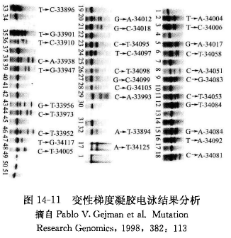
3. Analysis of the results of denaturing gradient gel electrophoresisAfter electrophoresis, EB staining or silver staining was used. The results were analyzed to determine whether the point mutation occurred. The results can be further analyzed by gene sequencing, as shown in Figure 14-11.
Caveat
① Since the use of the "GC-clamp" technique allows most of the mutations to fall into the low-temperature unchaining region, the end of the "GC-clamp" connection is also very important, which directly affects the effectiveness of the assay, so it is very important to select the optimal primer position by using a computer program. Some instruments sold by instrument companies have primer analysis software, which allows you to obtain the desired primer by entering the desired sequence.② When PCR products are analyzed by denaturing gradient gel electrophoresis, the mutation can be detected in 100% of the fragments up to 1 kb if the mutation occurs in the region of the DNA that is first unstranded, optimally in the range of 100-500 bp, and the mutant molecules can be detected in fragments up to 3,000 bp long if they are separated by constant denaturant capillary electrophoresis. Depending on the conditions in the laboratory, the appropriate assay should be selected. This can be carried out by first amplifying a portion of a large fragment by PCR, followed by superimposed screening.③ Amplify the genomic DNA with two primers, with the 5' end of one primer linked to a 40-45 bp GC-rich sequence. Because a heteroduplex must be formed to ensure a detection rate close to 100%, an equal amount of normal DNA molecules must be added to form the heteroduplex before analysis because mutant heterozygotes may be formed.④ When applying the "GC-clamp" denaturing gradient gel electrophoresis technique to study certain genes, if the gene contains a high level of GC bases (70%), it is not suitable to screen by DGGE because the CpG region is a hotspot of mutation, so it is necessary to screen a large number of genes by single-stranded conformational polymorphism technique. It is more difficult to find the exact cause of the false-negative problems encountered with heterologous double-stranded denaturing gradient gel electrophoresis analysis with GC-clamp, and it should be noted that methylated bases may be detected. So in the end sequencing must be performed to determine if what is detected is a true mutant base.⑤ DGGE and TGGE have commercialized electrophoresis devices, and once established, the method is easier to operate and is suitable for the detection and screening of large samples.
For more product details, please visit Aladdin Scientific website.