
Photodynamic Therapy Technology: Mechanisms of Action, Photosensitizer Systems, and Experimental Evaluation
Photodynamic Therapy Technology: Mechanisms of Action, Photosensitizer Systems, and Experimental Evaluation
Photodynamic therapy (PDT) is a photochemical treatment technology jointly driven by photosensitizers, light of a specific wavelength, and molecular oxygen. After photosensitizers accumulate in target cells or diseased tissues, they are activated by light irradiation to generate singlet oxygen, free radicals, and other reactive oxygen species, which further induce organelle damage, membrane lipid oxidation, vascular disruption, and local immune responses. Because the irradiation range and photosensitizer distribution are relatively controllable, PDT has important application value in tumors, hyperproliferative skin diseases, HPV-related lesions, vascular diseases, and antimicrobial therapy.
Keywords: photodynamic therapy; PDT; photosensitizer; singlet oxygen; reactive oxygen species; irradiation parameters; tumor therapy; combination therapy
1 Basic Concepts of Photodynamic Therapy
1.1 Technical Scope of PDT
PDT is a local treatment technology based on photosensitization reactions. Its effect is not determined by the drug alone, but by photosensitizer distribution, irradiation range, light source parameters, and tissue oxygen supply. Without effective light irradiation, photosensitizers usually show relatively low dark toxicity. After exposure to light at a matching wavelength, they can enter an excited state and generate reactive oxygen species, causing oxidative damage to diseased cells, vascular endothelial cells, or microbial structures within the irradiated area.
1.2 Three-Component Reaction System
(1) Photosensitizer
The photosensitizer absorbs light energy and enters an excited state. Its absorption peak, molar extinction coefficient, reactive oxygen species yield, dark toxicity, hydrophilicity/hydrophobicity, subcellular localization, and tissue clearance rate all affect PDT efficacy. In vitro experiments focus more on cellular uptake, photostability, and subcellular localization. In vivo experiments also need to consider tissue distribution, metabolic clearance, and the duration of photosensitivity.
(2) Excitation light
The excitation light must match the absorption peak of the photosensitizer. Wavelength determines excitation efficiency and tissue penetration depth. Power density and energy density determine energy input per unit time and total light dose. The irradiation mode affects photobleaching, oxygen depletion, and the risk of local thermal effects. When the wavelength does not match, even increasing irradiation time may not produce a stable photodynamic effect.
(3) Oxygen
Oxygen participates in the generation of singlet oxygen and some free radicals and is an important substrate in PDT reactions. Tumor hypoxia, insufficient blood perfusion, or rapid oxygen consumption caused by high-intensity continuous irradiation may reduce ROS generation efficiency. In hypoxia-related models, oxygen supply status should be analyzed together with photosensitizer uptake, light dose, and mode of cell death.
1.3 Technical Characteristics
The characteristics of PDT are mainly reflected in local activation, selective oxidative damage, and multi-target effects. Local activation arises from control of the irradiation range, selective damage comes from relative enrichment of photosensitizers in diseased tissues and restriction of the irradiated area, and multi-target effects are manifested as organelle damage, membrane lipid oxidation, vascular occlusion, inflammatory responses, and immune regulation. Compared with thermal ablation, chemotherapy, or radiotherapy, PDT does not primarily rely on thermal injury, systemic drug exposure, or ionizing radiation-induced DNA damage. Instead, it triggers local biological effects through photochemical reactions.
2 Photochemical Reaction Basis of PDT
2.1 Photosensitizer Excitation Process
After absorbing photons at a matching wavelength, a photosensitizer transitions from the ground state to an excited singlet state. The excited singlet state can release energy through fluorescence or undergo intersystem crossing to become a longer-lived excited triplet state. The triplet-state photosensitizer is the key intermediate for reactive oxygen species generation and can induce biological damage through type I or type II reactions.
(1) Type I reaction
Type I reactions are mainly based on electron transfer or hydrogen transfer and can generate reactive species such as superoxide anions, hydroxyl radicals, lipid radicals, and peroxides. This reaction pathway has certain advantages in hypoxic environments and is commonly used in hypoxic tumor PDT, novel photosensitizer design, and studies combining PDT with ferroptosis.
(2) Type II reaction
Type II reactions are mainly based on energy transfer. The excited-state photosensitizer transfers energy to triplet oxygen to generate singlet oxygen. Singlet oxygen is highly reactive, short-lived, and has a limited diffusion distance. It usually preferentially damages membrane lipids, proteins, and organelle structures near the photosensitizer and is one of the most important photochemical pathways in classical PDT.
(3) Factors affecting reaction proportions
Type I and type II reactions often coexist in actual PDT systems. Photosensitizer structure, oxygen concentration, substrate environment, irradiation intensity, and microenvironmental redox state all affect the relative proportions of these two reaction types. Hypoxic environments, electron donor-rich environments, or specific photosensitizer structures may increase the proportion of type I reactions. When oxygen supply is sufficient and the triplet-state energy transfer efficiency of the photosensitizer is high, type II reactions are usually more prominent.
2.2 Targets of Reactive Oxygen Species Damage
Reactive oxygen species generated by PDT have a short reaction radius and short lifetime, so they usually preferentially attack biomolecules near the photosensitizer. Different subcellular localizations lead to different damage outcomes.
(1) Mitochondrial damage
Mitochondrial localization or damage near mitochondria can lead to decreased membrane potential, reduced ATP, cytochrome c release, and activation of the caspase cascade. Annexin V/PI, JC-1, Caspase-3, and PARP cleavage can be combined to assess the apoptotic process.
(2) Lysosomal damage
Lysosome-localized photosensitizers can cause disruption of acidic compartments, cathepsin release, and changes in autophagic flux. LC3-II, p62, LysoTracker, and lysosomal membrane permeability assays can be used to distinguish protective autophagy from death-related autophagy.
(3) Membrane lipid oxidation
When photosensitizers localize near the plasma membrane or organelle membranes, PDT can induce lipid peroxidation and loss of membrane integrity. C11-BODIPY, MDA, 4-HNE, PI staining, and LDH release can be used to evaluate membrane oxidative damage and necrosis-like cell death.
(4) Vascular and matrix damage
If photosensitizers are mainly distributed in vascular endothelium or extracellular matrix, PDT effects may manifest as increased vascular permeability, thrombosis, decreased perfusion, and local hypoxia changes. In vivo models can be evaluated using CD31, perfusion imaging, and hypoxia probes.
3 Photosensitizer Systems
3.1 Key Points in Photosensitizer Selection
The photosensitizer is the core component of a PDT system, and its properties directly affect photoreaction efficiency and biological effects. An ideal photosensitizer should have a defined absorption peak, high reactive oxygen species yield, low dark toxicity, good photostability, and interpretable cellular or tissue distribution characteristics.
(1) Spectral compatibility
The absorption peak of the photosensitizer should match the wavelength of the experimental light source. If the light source wavelength deviates from the absorption peak, photosensitizer excitation efficiency decreases, and experiments may show weak ROS signals, unstable phototoxicity, or a requirement for excessively high light doses.
(2) Dark toxicity and phototoxicity window
Photosensitizers should maintain low toxicity under dark conditions and produce a clear phototoxic response after irradiation. When dark toxicity is too high, cell death is difficult to attribute to a photodynamic reaction. When phototoxicity is insufficient, the causes should be investigated from aspects such as cellular uptake, light dose, ROS generation, and oxygen supply.
(3) Cellular uptake and subcellular localization
Cellular uptake affects the usable concentration of a photosensitizer, while subcellular localization determines the initial damage target. Different localization patterns, such as mitochondria, lysosomes, plasma membrane, endoplasmic reticulum, or extracellular matrix localization, lead to different modes of cell death and molecular indicators after PDT.
(4) In vivo distribution and clearance
In vivo PDT also requires attention to photosensitizer distribution in tumors, skin, liver, spleen, blood vessels, and normal tissues. Slow clearance may increase the risk of prolonged photosensitivity, whereas rapid clearance may shorten the effective treatment window.
3.2 Main Types of Photosensitizers
Different photosensitizers should not simply be listed by name. They should be selected based on “spectral matching—tissue/cellular uptake—ROS type—experimental use.”
(1) 5-ALA prodrug system
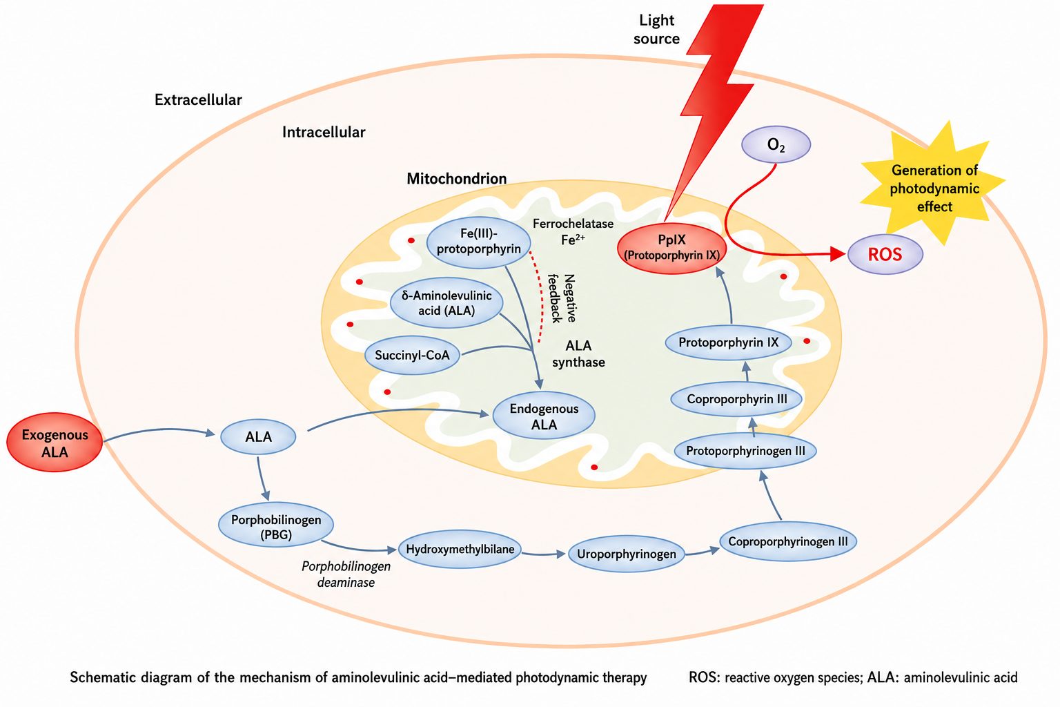
5-ALA promotes intracellular accumulation of protoporphyrin IX (PpIX) through the heme biosynthesis pathway and is suitable for skin, mucosal, HPV-related lesion, and epithelial proliferation models. The experimental focus of this system is not simply comparing 5-ALA concentrations, but evaluating PpIX fluorescence accumulation, incubation time, cellular metabolic status, and ROS generation capacity after irradiation. For PDT studies related to cervical lesions, skin lesions, or condyloma acuminatum, the 5-ALA system better reflects the relationship between metabolic differences in diseased cells and local phototoxicity.
(2) Porphyrin and chlorin photosensitizers
Porphyrin photosensitizers are suitable for establishing classical PDT mechanism models and can be used to analyze singlet oxygen generation, mitochondrial damage, apoptosis, and vascular effects. Chlorin photosensitizers usually absorb at longer wavelengths and are suitable for tumor model research under red or near-red light excitation. The focus of these photosensitizers should be placed on absorption peak and light source compatibility, cellular uptake efficiency, subcellular localization, and tissue retention time in vivo, rather than evaluating quality only by cell killing rate.
(3) Phthalocyanine and near-infrared-responsive photosensitizers
Phthalocyanine and near-infrared-responsive photosensitizers are suitable for deep tissue, three-dimensional tumor spheroids, organoids, or in vivo solid tumor models. Their research focus lies in long-wavelength excitation, tissue penetration, hypoxia adaptation, and compatibility with nanodelivery systems. If photothermal effects also exist, temperature monitoring and photothermal controls should be included to avoid misinterpreting thermal injury as PDT effects.
(4) Antimicrobial and mechanistic control photosensitizers
Methylene blue, toluidine blue, rose bengal, and similar compounds are commonly used for antimicrobial PDT, biofilm disruption, and in vitro ROS mechanism studies. These systems are more suitable for verifying singlet oxygen, free radical, or membrane damage processes and should not be directly extrapolated to deep-tissue tumor PDT. Experiments should carefully record irradiation wavelength, microbial uptake, biofilm penetration, and dark toxicity background.
4 Irradiation Parameters and Oxygen Supply Control
4.1 Light Source Parameters
PDT light source parameters include wavelength, power density, energy density, irradiation time, irradiation distance, spot area, and light source type. Wavelength determines whether the photosensitizer can be effectively excited. Power density determines energy input per unit time. Energy density represents the total light dose. Irradiation time and mode affect the risks of photobleaching, oxygen depletion, and local thermal effects.
Table 1 Key points for recording PDT irradiation parameters
Parameter | Recording content | Experimental significance |
Wavelength | nm value and spectral range | Determines photosensitizer excitation efficiency |
Power density | mW/cm² | Affects ROS generation rate and risk of thermal effects |
Energy density | J/cm² | Represents total light dose |
Irradiation time | Continuous or intermittent irradiation | Affects oxygen depletion and photobleaching |
Light source type | Laser, LED, semiconductor light source | Affects monochromaticity, uniformity, and operability |
Irradiation distance | Distance from light source to sample | Affects the actual light dose reaching the sample |
Spot area | Actual irradiated area | Determines treatment range and dose uniformity |
Temperature monitoring | Temperature changes before and after irradiation | Used to exclude interference from photothermal effects |
4.2 Oxygen Supply Limitation
PDT reactions continuously consume oxygen, and hypoxia is an important factor limiting PDT efficacy. Tumor tissues often have insufficient perfusion and hypoxic regions, and high-intensity continuous irradiation may further accelerate local oxygen depletion, reducing ROS generation.
(1) Optimization of irradiation strategy
Intermittent irradiation or low-power irradiation can slow local oxygen depletion and allow oxygen time to diffuse back into the reaction area. This strategy is suitable for three-dimensional tumor spheroids, organoids, or solid tumor models with obvious oxygen supply limitations.
(2) Optimization of oxygen supply systems
Oxygen-carrying materials, catalytic oxygen-generating systems, and microenvironment-responsive delivery systems can improve local oxygen supply. When evaluating such systems, oxygen level, ROS generation, and mode of cell death should be measured simultaneously, rather than judging oxygen supply effects only by changes in cell viability.
(3) Optimization of photosensitizer pathways
Photosensitizers with type I reaction advantages can maintain a certain ability to generate free radicals under hypoxic conditions. In hypoxic PDT studies, singlet oxygen-dominated effects should be distinguished from free radical-dominated effects and analyzed together with HIF-1α, oxygen probes, and ROS type detection.
5 Biological Effects of PDT
5.1 Modes of Cell Death
PDT can induce apoptosis, necrosis, autophagy alteration, and mixed forms of cell death. Low to moderate oxidative damage is often associated with decreased mitochondrial membrane potential, caspase activation, and apoptosis. Strong membrane damage or rapid lipid peroxidation can cause necrosis-like cell death. Lysosomal damage may be accompanied by disruption of acidic compartments and altered autophagic flux. PDT evaluation should not rely only on cell viability readouts; membrane integrity, mitochondrial function, apoptotic markers, lipid peroxidation, and cellular morphological changes should also be assessed comprehensively.
5.2 Vascular and Immune Effects
The tissue-level effects of PDT do not only arise from direct death of diseased cells, but also include vascular damage and changes in the immune microenvironment. After vascular endothelial injury, increased permeability, thrombosis, decreased perfusion, and secondary hypoxia may occur. Damage-associated molecules released by oxidative injury can promote antigen exposure, inflammatory cytokine release, and immune cell recruitment. In tumor PDT research, CD31, perfusion imaging, hypoxia probes, CRT exposure, HMGB1 release, ATP release, and CD8+ T cell infiltration can be combined to distinguish the contributions of direct phototoxicity, vascular effects, and immune effects to the overall response.
6 Disease Models and Application Research
6.1 Tumor PDT Models
Tumor PDT studies can select two-dimensional cells, three-dimensional tumor spheroids, organoids, xenografts, or orthotopic models according to the complexity of the research question.
(1) Two-dimensional cell models
Two-dimensional cell models are suitable for rapid screening of photosensitizer concentration, incubation time, dark toxicity, phototoxicity, and irradiation parameters. This model is easy to operate and has good reproducibility, but it cannot fully reflect limitations in light penetration, oxygen diffusion, and drug distribution in solid tissues.
(2) Three-dimensional tumor spheroids and organoid models
Three-dimensional tumor spheroids can simulate light attenuation, hypoxia gradients, and insufficient drug penetration, making them suitable for evaluating PDT penetration depth and spatial distribution of cell death. Organoid models are closer to tissue heterogeneity and can be used to analyze the sensitivity of different cell populations to photodynamic damage.
(3) Xenograft and orthotopic models
Animal models can be used to evaluate the effects of PDT on tumor vasculature, immune microenvironment, tissue necrosis boundaries, and systemic safety. At this stage, tumor volume change alone should not be the only observation. Photosensitizer tissue distribution, ROS-related damage, vascular markers, immune infiltration, and recurrence should be analyzed comprehensively.
6.2 Skin, Mucosal, and HPV-Related Models
Skin and mucosal lesions are superficial, accessible to local administration, and easy to irradiate, making them suitable for ALA-PDT and local PDT studies. HPV-related lesion models can be studied around PpIX accumulation, E6/E7 expression, abnormal p16/Ki-67 co-expression, epithelial cell proliferation, and local inflammatory changes. In PDT studies related to cervical lesions, in addition to abnormal cell clearance, epithelial structure preservation, inflammatory microenvironment regulation, and tissue repair quality should also be considered.
6.3 Antimicrobial and Vascular Lesion Models
(1) Antimicrobial PDT models
Antimicrobial PDT mainly focuses on bacterial inactivation, biofilm disruption, and local infection control. Common evaluation indicators include CFU counts, biofilm staining, ROS detection, bacterial membrane integrity, and recurrent growth.
(2) Vascular lesion models
PDT for vascular lesions focuses more on photosensitizer distribution in vascular endothelium, endothelial damage after irradiation, vascular occlusion, and normal tissue protection. Endothelial cell viability, CD31 staining, blood flow perfusion, and tissue structure observation can be used to evaluate vascular-selective damage.
7 Combination Therapy and Technical Expansion
7.1 PDT and Immunotherapy
PDT can induce immunogenic cell death and promote antigen exposure and T cell responses. When combined with immune checkpoint inhibitors, tumor vaccines, or immune adjuvants, the analysis should focus on CRT exposure, HMGB1 release, ATP release, dendritic cell maturation, CD8+ T cell infiltration, and PD-L1 expression changes. If responses occur in distant non-irradiated lesions, systemic antitumor immune effects can be further evaluated.
7.2 PDT and Ferroptosis
PDT-induced ROS and lipid peroxidation can intersect with ferroptosis mechanisms. In combination studies, C11-BODIPY lipid ROS, GPX4, SLC7A11, GSH, and iron ion levels should be measured, and ferroptosis inhibitors or lipid peroxidation inhibitors should be used to verify mechanistic involvement. Observing enhanced cell death alone is insufficient to prove ferroptosis participation.
7.3 Nanodelivery and Responsive Design
Nanodelivery systems can improve photosensitizer water solubility, circulation stability, tissue enrichment, and cellular uptake. Common carriers include liposomes, polymer nanoparticles, metal-organic frameworks, albumin nanoparticles, and silica nanoparticles. Responsive designs can use pH, enzymes, reducing environments, hypoxia, or light irradiation to trigger release, enabling more effective photosensitizer enrichment in the diseased microenvironment.
(1) Delivery performance evaluation
Nano-PDT systems should be evaluated for particle size, PDI, zeta potential, drug loading, encapsulation efficiency, release profile, and serum stability. If these basic parameters are unstable, efficacy differences in subsequent cell or animal experiments are difficult to interpret.
(2) Cellular and tissue uptake evaluation
Delivery systems should further evaluate cellular uptake efficiency, subcellular localization, tumor tissue enrichment, and normal tissue distribution. Targeted delivery systems should also include blank carrier, non-targeted carrier, and competitive inhibition controls.
(3) Phototoxicity and safety evaluation
Nanocarriers themselves may have cytotoxicity or immunostimulatory effects, so PDT-induced cell death rate should not be the only comparison. Dark toxicity, carrier toxicity after irradiation, blood compatibility, and safety in major organs in vivo should all be included in the evaluation system.
8 Experimental Design and Result Evaluation
8.1 Control Setup
PDT experiments should include at least a blank control, light-only control, photosensitizer dark-treatment group, and PDT treatment group. When solvents, nanocarriers, targeting ligands, or combined drugs are used, solvent controls, blank carrier controls, carrier plus irradiation controls, and combination-treatment controls should also be included. Without light-only controls and dark toxicity controls, it is impossible to distinguish photosensitizer toxicity, light-induced damage, and true photodynamic effects.
8.2 Result Evaluation
Evaluation of PDT results should cover the photosensitizer, irradiation, ROS, and biological endpoints. At the photosensitizer level, uptake, localization, and dark toxicity should be measured. At the irradiation level, wavelength, power density, energy density, and temperature changes should be recorded. At the ROS level, singlet oxygen, free radicals, and lipid peroxidation should be distinguished. At the biological endpoint level, modes of cell death, organelle damage, vascular effects, immune responses, and tissue repair status should be analyzed together. Early screening focuses on the phototoxicity window, mechanistic studies focus on ROS type and death pathways, and in vivo application studies focus on tissue distribution, efficacy boundaries, and safety.
9 Related Reagent and Material Selection
Table 2 Photosensitizers and ROS detection reagents for photodynamic therapy research
Cat. No. | Product Name | Specification/Purity | Application Module | Application Positioning |
5-Aminolevulinic acid hydrochloride | ≥99% | ALA-PDT prodrug system | Induces intracellular PpIX accumulation; used in skin, mucosal, and HPV-related PDT studies | |
5-Aminolevulinic acid HCl | 10mM in DMSO | ALA-PDT prodrug system | Used for ALA-PDT cell experiments and phototoxicity condition screening | |
Protoporphyrin IX | Moligand™,10mM in DMSO | Porphyrin photosensitizer | Used for PpIX-related photosensitization reactions and cellular uptake evaluation | |
Chlorin e6 | Moligand™, 10 mM in DMSO | Chlorin photosensitizer | Used in red-light PDT, tumor models, and nanodelivery system research | |
CHLORIN E6(CE6) | Moligand™,≥90% | Chlorin photosensitizer | Used for Ce6-PDT cellular phototoxicity and ROS generation studies | |
Methylene blue | 10mM in DMSO | Phenothiazine photosensitizer | Used for antimicrobial PDT, ROS generation, and photosensitization evaluation | |
Methylene blue | 0.05% | Phenothiazine photosensitizer | Used for antimicrobial PDT and staining/photosensitization treatment systems | |
Methylene blue | 0.1% | Phenothiazine photosensitizer | Used for antimicrobial PDT or cellular phototoxicity studies under different concentration conditions | |
Methylene blue | ≥70% | Phenothiazine photosensitizer | Used for basic photosensitization reactions and antimicrobial PDT experiments | |
Methylene blue | 1% | Phenothiazine photosensitizer | Used for higher-concentration staining or local treatment systems | |
Rose bengal | ≥95% | Singlet oxygen photosensitizer | Used for singlet oxygen generation, antimicrobial PDT, and photochemical mechanism controls | |
Rose bengal | Biological Stain | Singlet oxygen photosensitizer | Used for biological staining, photosensitization reactions, and antimicrobial PDT research | |
Rose bengal | ≥90% | Singlet oxygen photosensitizer | Used for photosensitization reactions, ROS generation, and mechanism controls | |
Indocyanine Green | Moligand™, 10mM in DMSO | Near-infrared responsive photosensitizer/imaging material | Used for near-infrared PDT, image guidance, and distinguishing photothermal effects | |
Indocyanine Green (ICG) | Moligand™, ≥75% | Near-infrared responsive photosensitizer/imaging material | Used for ICG-related phototherapy, delivery, and in vivo imaging research | |
2',7'-Dichlorodihydrofluorescein diacetate | 10mM in DMSO | Total ROS detection | Used to detect intracellular total ROS changes after PDT | |
2',7'-Dichlorodihydrofluorescein diacetate(DCFH-DA) | ≥97% | Total ROS detection | Used for evaluating intracellular ROS generation and analyzing phototoxicity mechanisms | |
1,3-Diphenylisobenzofuran | ≥97% | Singlet oxygen detection | Used for singlet oxygen generation and photochemical reaction validation in solution systems | |
Singlet Oxygen Sensor Green Reagent |
| Singlet oxygen detection | Used to evaluate singlet oxygen generation in type II PDT reactions | |
ROS fluorescent probe DHE |
| ROS detection | Used to detect superoxide anion-related ROS changes |
Table 3 Lipid oxidation and organelle fluorescent probes for photodynamic therapy research
Cat. No. | Product Name | Specification/Purity | Application Module | Application Positioning |
1,3,5,7-Tetramethyl-2,6-diiodo-C3-SE-BODIPYs |
| BODIPY photosensitizer | Research on iodinated BODIPY photosensitizer systems | |
2,6-Diiodo-BODIPY 493/503 |
| BODIPY photosensitizer | Research on iodinated BODIPY photoreactions | |
BODIPY 650/665 NHS ester |
| Long-wavelength fluorescent labeling | Labeling of photosensitizer carriers, proteins, or nanodelivery systems | |
BODIPY 665/676 | ≥99% | Long-wavelength responsive fluorescence system | Long-wavelength excitation imaging or delivery tracking | |
BODIPY 581/591 C11 | ≥99% | Lipid peroxidation detection | Detection of PDT-induced lipid ROS | |
BODIPY-581/591 NHS ester | ≥98% | Lipid oxidation/fluorescent labeling | Derivatized labeling related to lipid peroxidation | |
BODIPY 500/510 C1, C12 | ≥99% | Membrane lipid and fatty acid probe | Observation of lipid uptake and membrane lipid environment | |
BODIPY 500/510 C1, C12 (Fatty Acid Green Fluorescence Probe) |
| Membrane lipid and fatty acid probe | Fatty acid uptake and membrane lipid analysis | |
BODIPY 505/515 | Moligand™, 10 mM in DMSO | Basic BODIPY fluorescent probe | Cellular uptake, carrier tracking, and fluorescent labeling | |
BODIPY 540 (purity 99%) | ≥99% | BODIPY fluorescent probe | Cellular imaging and photosensitizer system labeling | |
BODIPY 558/568 C12 | ≥98% | Lipid/membrane-related probe | Observation of membrane lipid environment and lipid distribution | |
BODIPY 564/570 |
| BODIPY fluorescent probe | Cellular imaging and delivery tracking | |
BODIPY 576/589 | ≥96% | BODIPY fluorescent probe | Fluorescent labeling and cellular imaging | |
BODIPY 576/589 | 10 mM in DMSO | BODIPY fluorescent probe | Fluorescent labeling and cellular imaging | |
JC-1 | A cationic, fluorescent, carbocyanine dye that can be used as a ratiometric indicator of mitochondrial potential δΨm in cells, tissues, and isolated mitochondria. | Mitochondrial function detection | Evaluation of mitochondrial membrane potential changes | |
JC-1 iodide | ≥95% | Mitochondrial function detection | Evaluation of mitochondrial membrane potential changes | |
JC-1 | ≥95% | Mitochondrial function detection | Evaluation of mitochondrial membrane potential changes | |
JC-10 | ≥95% | Mitochondrial function detection | Evaluation of mitochondrial membrane potential changes | |
Mito-Tracker Far-Red | BioReagent, ≥95% | Mitochondrial localization observation | Analysis of photosensitizer subcellular localization and mitochondrial status | |
Mito-Tracker Deep Red FM |
| Mitochondrial localization observation | Mitochondrial labeling and localization analysis | |
LysoTracker Blue DND-22 |
| Lysosome observation | Lysosomal localization and changes in acidic compartments | |
LysoTracker Red | ≥97% | Lysosome observation | Lysosomal integrity and changes in acidic compartments | |
LysoTracker Yellow HCK 123 | ≥98% | Lysosome observation | Lysosomal localization and damage analysis | |
Lyso-Tracker Red |
| Lysosome observation | Lysosomal structure and acidic compartment observation | |
Fluolysotm red (lysosomal red fluorescent probe) |
| Lysosome observation | Lysosomal localization and damage evaluation | |
Fluolysotm deep red (lysosomal dark red fluorescent probe) |
| Lysosome observation | Lysosomal localization and multichannel imaging | |
ER-Tracker Green |
| Endoplasmic reticulum observation | ER localization and stress-related observation | |
ER-Tracker Red |
| Endoplasmic reticulum observation | ER localization and stress-related observation | |
ER-Tracker Red |
| Endoplasmic reticulum observation | ER localization and stress-related observation |
10 Frequently Asked Questions
10.1 Why must dark toxicity controls be included in PDT experiments?
Photosensitizers themselves may affect cell viability, mitochondrial function, or membrane structure. Dark toxicity controls help determine whether cell damage comes from the photosensitizer itself rather than from the photodynamic reaction after light activation.
10.2 Can the light source wavelength be replaced arbitrarily?
No. The light source wavelength must match the absorption peak of the photosensitizer. A wavelength deviating from the absorption peak reduces excitation efficiency, and increasing the light dose may still fail to produce the same PDT effect.
10.3 Can a single ROS probe prove the PDT mechanism?
No. Different ROS probes vary in sensitivity to oxidative species, irradiation conditions, and photosensitizer interference. Mechanistic studies should combine ROS probes, scavengers, lipid peroxidation indicators, and cell death pathway analysis.
10.4 Why may in vitro PDT results not translate directly to in vivo conditions?
In vivo systems have limitations in light penetration, oxygen diffusion, vascular perfusion differences, uneven drug distribution, and effects of the immune microenvironment. Two-dimensional cell experiments are suitable for parameter screening, but they cannot replace validation in three-dimensional models and animal models.
10.5 What indicators should be measured in studies combining PDT with ferroptosis?
Lipid ROS, GPX4, SLC7A11, GSH, and iron ion levels should be measured, and ferroptosis inhibitors or lipid peroxidation inhibitors should be used for validation. Observing enhanced cell death alone is insufficient to prove ferroptosis involvement.
The technical value of photodynamic therapy comes from the coordinated control of photosensitizer, irradiation, and oxygen supply. By rationally selecting photosensitizer systems, standardizing irradiation parameters, distinguishing ROS types, and establishing multilevel evaluation indicators, PDT-induced local damage mechanisms and differences in therapeutic response can be interpreted more accurately.