Hoechst 33258 staining solution - BioReagent, Biological Stain, Biological dye grade, Suitable for Immunofluorescence(IF), for fluorescence analysis, for cell culture, for microscopy, ready-to-use, 1.0 mg/mL in H₂O , CAS No.23491-45-4

CAS: 23491-45-4 Cat. No.: H1373484 Molecular Weight: 533.90 Beilstein Registry Number: 4088183 EC Number: 245-690-6 PubChem CID: 31953
AVAILABLE TO ORDER
GRADE & PURITY BioReagent ? BioReagent grade — tested suitable for life-science and molecular-biology use. Use for cell culture, assays, and biochemical work needing biological compatibility. Ready-to-use ? Ready-to-use — supplied pre-formulated at working concentration, no prep needed. Use to save time and reduce pipetting/dilution errors. Biological Stain ? Biological stain grade — dyes characterized for staining cells and tissues. Use in histology and microscopy where staining consistency matters. for Fluorescence analysis ? Fluorescence-analysis grade — very low fluorescent impurities for clean spectra. Use in fluorescence assays where background interferes. Biological dye grade ? Biological stain grade — dyes characterized for staining cells and tissues. Use in histology and microscopy where staining consistency matters. for Microscopy ? Microscopy grade — reagents/stains suited to sample prep and imaging. Use in microscopy where clarity and low background are needed. for Cell culture ? Cell-culture grade — low endotoxin and contaminants to support viable cell growth. Use in mammalian/other cell culture media and supplements. Suitable for Immunofluorescence(IF) ? IF grade — validated for immunofluorescence with low background staining. Use to localize targets in cells/tissue by fluorescence microscopy. 1.0 mg/mL in H₂O
Synonyms
Hoechst 33258 | Hoechst 33258 Stain Solution | Hoechst 33258 Stain Solution(1mg/ml) | Hoechst 33258 Staining Dye Solution | Hoechst 33258 Solution (1 mg/mL)
Storage
Protected from light,Store at -20°C,Avoid repeated freezing and thawing
Shipped In
Ice chest + Ice pads
Application
Cell Apoptosis Analysis, Cell Cycle Analysis, Cell Stain, Flow Cytometry, IF/ICC
 ·  off list, applied to all prices below.
Size
Status
Price
Qty
1ml
H1373484-1ml
2
$24.90
10×1ml
H1373484-10×1ml
1-2 wks(?)
Item is derived from our semi-finished stock and is processed in 1-2 weeks.
$139.90
Enter a quantity for the sizes you want to add.
🧪

Why this grade

BioReagent, Biological Stain, Biological dye grade, Suitable for Immunofluorescence(IF), for fluorescence analysis, for cell culture, for microscopy, ready-to-use, 1.0 mg/mL in H₂O Biological dye grade,Biological Stain,BioReagent,for Cell culture,for Fluorescence analysis,for Microscopy,Ready-to-use,Suitable for Immunofluorescence(IF) for sensitive chromatographic and analytical workflows requiring minimal baseline interference.

🌡

Storage & shipping

Protected from light,Store at -20°C,Avoid repeated freezing and thawing Ships Ice chest + Ice pads Check lot-specific COA for exact specifications.

📋

Quality documents

SDS, COA, datasheet, and spec sheet available for download. Lot-specific COA accessible via lot number lookup.

📚

Literature proof

Cited in 33 peer-reviewed publications across chromatography, organic synthesis, and cross-coupling reactions.

Overview

Hoechst 33258 is a blue to blue-green fluorescent live cell dye that can label DNA. Hoechst 33258 can specifically bind to the DNA groove (tending to bind to DNA rich in A/T at the same time), resulting in a significant increase in fluorescence intensity. Hoechst 33258 can pass through the cell membrane, causing changes in DNA structure, such as G2/M phase arrest. Hoechst 33258 can bind to live cells or fixed cells, and the fluorescence intensity increases with the increase of solution pH. As a DNA-specific probe, Hoechst 33258 can be used for detecting DNA content and analyzing cell cycle, etc. The excitation wavelength of Hoechst 33258 is 350-365 nm, and the emission wavelength is 460-490 nm.

The concentration of this product is 1.0 mg/mL. When used for nuclear staining, the recommended working concentration is 1-10 μg/mL.

Instruction for use 

1.For fixed cells or tissues:

a. The cells were inoculated onto the culture plates/flasks and cultured until they reached the logarithmic growth phase. If cell fixation was required, the cells were fixed with 4% paraformaldehyde at room temperature for 10-15 minutes, then washed twice with PBS, each time for 5 minutes.

b. Add the Hoechst 33258 working solution and incubate at 37°C in the dark for 10-30 minutes.

c. Remove the Hoechst 33258 staining solution and wash the cells twice with PBS for 5 minutes each time.

2.For living cells or tissues:

a. Collect the cells, centrifuge at 1000 rpm for 5 minutes, and discard the supernatant.

b. Resuspend the cells in PBS, add the Hoechst 33258 working solution, and incubate at room temperature in the dark for 15-20 minutes.

c. Centrifuge at 1000 rpm for 5 minutes, discard the staining solution, wash twice with PBS for 5 minutes each time; then resuspend the cells in PBS.

d.Observe directly under a fluorescence microscope or under a fluorescence microscope after mounting. When cells undergo apoptosis, the nuclei of apoptotic cells will show dense and compact staining, or fragmented and dense staining.

Matters needing attention

1.Depending on the application of Hoechst 33258 staining solution, it is recommended to perform a titration before use to determine the optimal working concentration.

2.Fluorescent dyes all have the problem of quenching. It is recommended to complete the detection as soon as possible after staining. Live cells or tissues should be observed immediately after staining.

3.This product is only for professional scientific research use. It shall not be used for clinical diagnosis or treatment, nor for food or medicine. It shall not be stored in ordinary residences.

4.For your safety and health, please wear laboratory coats and disposable gloves during operation.

Specifications

Synonyms
Hoechst 33258 | Hoechst 33258 Stain Solution | Hoechst 33258 Stain Solution(1mg/ml) | Hoechst 33258 Staining Dye Solution | Hoechst 33258 Solution (1 mg/mL)
Specifications & Purity
BioReagent, Biological Stain, Biological dye grade, Suitable for Immunofluorescence(IF), for fluorescence analysis, for cell culture, for microscopy, ready-to-use, 1.0 mg/mL in H₂O
Stability And Storage
Store at -20℃ long term (12 months). Store in the dark. Avoid repeated freezing and thawing.
Storage
Protected from light, Store at -20°C, Avoid repeated freezing and thawing
Shipped In
Ice chest + Ice pads
This product requires cold chain shipping. Ground and other economy services are not available.
Grade
Biological dye grade, Biological Stain, BioReagent, for Cell culture, for Fluorescence analysis, for Microscopy, Ready-to-use, Suitable for Immunofluorescence(IF)
Images
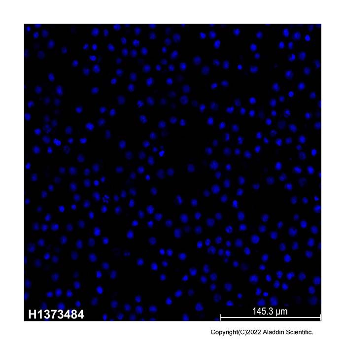
Hoechst 33258 Staining Solution (H1373484) - Cell Staining 
Apoptosis was induced in Jurkat cells by a 16-hour treatment with 2 μM camptothecin (C111281) and assessed using Hoechst 33258 staining solution (H1373484). This dye exhibits enhanced binding to condensed and fragmented chromatin in apoptotic cells, resulting in intense blue fluorescence under confocal laser scanning microscopy. In contrast, viable cells show only weak, diffuse nuclear fluorescence.
Names and Identifiers
Isomeric SMILES CN1CCN(CC1)C2=CC3=C(C=C2)N=C(N3)C4=CC5=C(C=C4)N=C(N5)C6=CC=C(C=C6)O.Cl.Cl.Cl
WGK Germany 3
RTECS SM1140500
PubChem CID 31953
Molecular Weight 533.90
Beilstein 4088183
Application
ApplicationDilution info
Flow Cytometry

1-10 μg/ml

Documentation

📋 Safety Data Sheet (SDS)

Comprehensive hazard, handling, storage, and regulatory compliance document.

Download SDS →

✅ Certificate of Analysis (COA)

Lot-specific quality data. Enter your lot number to retrieve the exact COA.

Look up COA →

📊 Datasheet

Quick-reference summary of product specifications and applications.

View datasheet →

🔬 Specification Sheet

Full quality attributes and acceptance criteria for this grade.

View spec sheet →

Advanced Data

Certificates(CoA,COO,BSE/TSE and Analysis Chart)
C of A & Other Certificates(BSE/TSE, COO):
Analytical Chart:
Chemical and Physical Properties
SensitivityLight-sensitive
Melt Point(°C)314°C
Documents & Articles
Suitable for Immunofluorescence(IF)
Ready-to-use Reagents
Biological Stain
Choosing Reagents ‘for Fluorescence Analysis’: A Practical Guide
For Cell Culture
Hoechst Staining for Apoptosis Detection: Experimental Workflow
Cell Live/Dead Staining and Apoptosis Detection Techniques
Principles and Methodological Framework for Nucleic Acid Staining and Organelle Labeling
Fluorescent Probes: A Complete Beginner’s Guide — Definitions & Emission Mechanisms, Signal Readouts, Classification Framework, Selection Roadmap, and Product Tables (Tables 1–4)
dsDNase-Mediated Hydrolysis of Double-Stranded DNA: Enzymatic Characteristics, Methodological Strategies, and Bioanalytical Applications
Physicochemical Properties of Diphenylamine, Analytical Applications, and a Review of Its DNA Colorimetric Reaction
Comparative Analysis and Selection Strategies for Mounting Media in Immunological Experiments
Cell Apoptosis Detection by TUNEL Labeling: Signal Interpretation and Result Evaluation
Principles, Applicable Scenarios, and Result Interpretation of Common Lipid Staining Methods
Types, Binding Mechanisms, and Experimental Selection of Nuclear Dyes
Practical Guide to TSA Tyramide Signal Amplification: How to Select Reagents, Optimize the Workflow, and Reduce Background When Detection Signals Are Weak for Low-Abundance Targets
Citations of This Product
References
1. Sijia Xiang, Zhuanghao Hou, Yu Wang, Yang Yang, Hongze Hu, Chang Yin, Guangming Huang, Kaiming Cao, Yangzhong Liu.  (2025)  Metal-induced nanoscale clusterization initiates protein liquid-liquid phase separation.  Cell Reports,      [PMID:40716064] [10.1016/j.celrep.2025.116067]
2. Meng Lan, Fansu Meng, Lanwen Gao, Anil K. Giri, Makiya Nishikawa, Kosuke Kusamori, Jaiwoo Lee, Mulazim Hussain Asim, Shumaila Arshad, Honghui Gu, Qi Li, Lina Yang, Zhong Chen, Zhenjiang Yang, Jiajia Qin, Yu Cai.  (2025)  Preparation of baicalin nano prodrug and its effect on inhibiting metastasis of triple-negative breast cancer.  Biomaterials Advances,      [PMID:40850054] [10.1016/j.bioadv.2025.214464]
3. Yaqian He, Zichuang Xu, Yaqian Yan, Jiajun Gao, Xuwu Zhang, Yuchu He, Qingzhi Luo, Desong Wang, Dawei Gao.  (2025)  ‘Pioneer’ T cells and ‘major force’ piezo-nanozymes synergize to achieve high-specificity targeted penetration and enhanced immunotherapy.  JOURNAL OF COLLOID AND INTERFACE SCIENCE,      [PMID:40966969] [10.1016/j.jcis.2025.139028]
4. Weiyuan Ma, Xiuwen Zhang, Le Zhuang.  (2023)  Exogenous Hydrogen Sulfide Induces A375 Melanoma Cell Apoptosis Through Overactivation of the Unfolded Protein Response.  Clinical Cosmetic and Investigational Dermatology,      [PMID:37396710] [10.2147/CCID.S412588]
5. Yingying Zhang, Tiange Wang, Yinping Zhuang, Tiandi He, Xiaoli Wu, Lin Su, Jun Kang, Jin Chang, Hanjie Wang.  (2021)  Sodium Alginate Hydrogel-Mediated Cancer Immunotherapy for Postoperative In Situ Recurrence and Metastasis.  ACS Biomaterials Science & Engineering,      [PMID:34757733] [10.1021/acsbiomaterials.1c01216]
6. Yunran Zhang, Haiping Zhou, Zhe Zhang, Yijun Zhu, Tao Wang, Liguo Yu, Hongrui Song.  (2020)  Redox/NIR dual-responsive PEG-betulinic acid/pluronic-cypate prodrug micelles for chemophotothermal therapy.  COLLOIDS AND SURFACES A-PHYSICOCHEMICAL AND ENGINEERING ASPECTS,      [PMID:] [10.1016/j.colsurfa.2020.125662]
7. Chenglong Ma, Wei Sun, Waqas Qamar Zaman, Zhenhua Zhou, Hao Zhang, Qicheng Shen, Limei Cao, Ji Yang.  (2020)  Lanthanides Regulated the Amorphization–Crystallization of IrO2 for Outstanding OER Performance.  ACS Applied Materials & Interfaces,      [PMID:32658446] [10.1021/acsami.0c08969]
8. Zhang Tao, Jiang Zhenqi, Chen Libin, Pan Chunshu, Sun Shan, Liu Chuang, Li Zihou, Ren Wenzhi, Wu Aiguo, Huang Pintong.  (2020)  PCN-Fe(III)-PTX nanoparticles for MRI guided high efficiency chemo-photodynamic therapy in pancreatic cancer through alleviating tumor hypoxia.  Nano Research,  13  (1): (273-281).  [PMID:] [10.1007/s12274-019-2610-6]
9. Jun Dai, Yinghao Li, Zi Long, Ruming Jiang, Zeyan Zhuang, Zhiming Wang, Zujin Zhao, Xiaoding Lou, Fan Xia, Ben Zhong Tang.  (2019)  Efficient Near-Infrared Photosensitizer with Aggregation-Induced Emission for Imaging-Guided Photodynamic Therapy in Multiple Xenograft Tumor Models.  ACS Nano,      [PMID:31820925] [10.1021/acsnano.9b07972]
10. Tao Zhang, Zhenqi Jiang, Ting Xve, Shan Sun, Juan Li, Wenzhi Ren, Aiguo Wu, Pintong Huang.  (2019)  One-pot synthesis of hollow PDA@DOX nanoparticles for ultrasound imaging and chemo-thermal therapy in breast cancer.  Nanoscale,  11  (45): (21759-21766).  [PMID:31482919] [10.1039/C9NR05671H]
11. Hailing Yang, Zhenjie Liu, Yonglong Song, Changjiang Hu.  (2019)  Hyaluronic acid-functionalized bilosomes for targeted delivery of tripterine to inflamed area with enhancive therapy on arthritis.  DRUG DELIVERY,      [PMID:31389248] [10.1080/10717544.2019.1636423]
12. Zigui Wang, Gaizhen Kuang, Zhiqiang Yu, Aimin Li, Dongfang Zhou, Yubin Huang.  (2019)  Light-activatable dual prodrug polymer nanoparticle for precise synergistic chemotherapy guided by drug-mediated computed tomography imaging.  Acta Biomaterialia,      [PMID:31128323] [10.1016/j.actbio.2019.05.047]
13. Zhao Shuai, Sun Shan, Jiang Kai, Wang Yuhui, Liu Yu, Wu Song, Li Zhongjun, Shu Qinghai, Lin Hengwei.  (2019)  In Situ Synthesis of Fluorescent Mesoporous Silica–Carbon Dot Nanohybrids Featuring Folate Receptor-Overexpressing Cancer Cell Targeting and Drug Delivery.  Nano-Micro Letters,  11  (1): (1-13).  [PMID:34137970] [10.1007/s40820-019-0263-3]
14. Rui Wang, Xing Hu, Junhui Pan, Guowen Zhang, Deming Gong.  (2019)  Interaction of isoeugenol with calf thymus DNA and its protective effect on DNA oxidative damage.  JOURNAL OF MOLECULAR LIQUIDS,      [PMID:] [10.1016/j.molliq.2019.03.018]
15. Jie Li, Suqin Shen, Fei Kong, Ting Jiang, Cui Tang, Chunhua Yin.  (2018)  Effects of pore size on in vitro and in vivo anticancer efficacies of mesoporous silica nanoparticles.  RSC Advances,  (43): (24633-24640).  [PMID:35539161] [10.1039/C8RA03914C]
16. Jie-Hua Shi, Yan-Yue Lou, Kai-Li Zhou, Dong-Qi Pan.  (2018)  Exploration of intermolecular interaction of calf thymus DNA with sulfosulfuron using multi-spectroscopic and molecular docking techniques.  SPECTROCHIMICA ACTA PART A-MOLECULAR AND BIOMOLECULAR SPECTROSCOPY,      [PMID:29935392] [10.1016/j.saa.2018.06.054]
17. Ying Wang, Yating Zhao, Yu Cui, Qinfu Zhao, Qiang Zhang, Sara Musetti, Karina A Kinghorn, Siling Wang.  (2017)  Overcoming multiple gastrointestinal barriers by bilayer modified hollow mesoporous silica nanocarriers.  Acta Biomaterialia,      [PMID:29037897] [10.1016/j.actbio.2017.10.025]
18. Huanan Wang, Wenqing Xu.  (2017)  Mito-methyl coumarin, a novel mitochondria-targeted drug with great antitumor potential was synthesized.  BIOCHEMICAL AND BIOPHYSICAL RESEARCH COMMUNICATIONS,      [PMID:28546001] [10.1016/j.bbrc.2017.05.116]
19. Hongxia Liu, Shuqin Wu, Jingmou Yu, Dun Fan, Jin Ren, Lei Zhang, Jianguo Zhao.  (2017)  Reduction-sensitive micelles self-assembled from amphiphilic chondroitin sulfate A-deoxycholic acid conjugate for triggered release of doxorubicin.  Materials Science & Engineering C-Materials for Biological Applications,      [PMID:28415498] [10.1016/j.msec.2017.02.030]
20. Huanan Wang, Ming Yao, Wenqing Xu.  (2016)  The antitumor effects of mitochondria-targeted 6-(nicotinamide) methyl coumarin.  Open Life Sciences,  11  (1): (542-551).  [PMID:] [10.1515/biol-2016-0070]
21. Haibo Mu, Qianjin Liu, Hong Niu, Dongdong Wang, Jiangjiang Tang, Jinyou Duan.  (2015)  Autophagy promotes DNA–protein crosslink clearance.  MUTATION RESEARCH-GENETIC TOXICOLOGY AND ENVIRONMENTAL MUTAGENESIS,      [PMID:26921017] [10.1016/j.mrgentox.2015.12.001]
22. Li Jingchao, Wang Tingting, Shi Yuanfei, Ye Zichen, Zhang Xun, Ming Jiang, Zhang Yafei, Hu Xinyan, Li Yun, Zhang Dongsheng, Xu Qianhe, Yang Jun, Chen Xiaolan, Liu Nian, Su Xinhui.  (2024)  A continuously efficient O2-supplying strategy for long-term modulation of hypoxic tumor microenvironment to enhance long-acting radionuclides internal therapy.  JOURNAL OF NANOBIOTECHNOLOGY,  22  (1): (1-15).  [PMID:38166931] [10.1186/s12951-023-02268-5]
23. Yanghui Chen, Yuan Li, Junliang Luo, Zixin Li, Yu Huang, Jia Cai, Dongneng Jiang, Defeng Zhang, Jichang Jian, Jun Qiang, Bei Wang.  (2024)  A novel study of brain microvascular endothelial cells induced by astrocyte conditioned medium for constructing blood brain barrier model in vitro: A promising tool for meningitis of teleost.  FISH & SHELLFISH IMMUNOLOGY,      [PMID:38266792] [10.1016/j.fsi.2024.109401]
24. Ouyang Boshu, Shan Caihua, Shen Shun, Dai Xinnan, Chen Qingwang, Su Xiaomin, Cao Yongbin, Qin Xifeng, He Ying, Wang Siyu, Xu Ruizhe, Hu Ruining, Shi Leming, Lu Tun, Yang Wuli, Peng Shaojun, Zhang Jun, Wang Jianxin, Li Dongsheng, Pang Zhiqing.  (2024)  AI-powered omics-based drug pair discovery for pyroptosis therapy targeting triple-negative breast cancer.  Nature Communications,  15  (1): (1-20).  [PMID:39215014] [10.1038/s41467-024-51980-9]
25. Zhang Hongmei, Yang Yunxia, Wang Yanqing.  (2025)  Comprehensive Investigations About the Binding Interactions of Sudan Dyes with DNA by Spectroscopy and Docking Methods.  JOURNAL OF FLUORESCENCE,      [PMID:39777621] [10.1007/s10895-024-04125-6]
26. Ziwei Yan, Hui Zhang, Jinghao Chen, Qingqing Xu, Shuaipeng Feng, Qinfu Zhao, Siling Wang.  (2024)  Cu(II)-doped mesoporous polydopamine as biodegradable nanoplatforms for photothermal-enhanced multi-mode anti-tumor therapy.  COLLOIDS AND SURFACES A-PHYSICOCHEMICAL AND ENGINEERING ASPECTS,      [PMID:] [10.1016/j.colsurfa.2024.133258]
27. Jingmou Yu, Yifei Zhang, Meilin Xu, Dengzhao Jiang, Wenbo Liu, Hongguang Jin, Pu Chen, Jing Xu, Lei Zhang.  (2024)  Innovative gelatin-based micelles with AS1411 aptamer targeting and reduction responsiveness for doxorubicin delivery in tumor therapy.  BIOMEDICINE & PHARMACOTHERAPY,      [PMID:38513599] [10.1016/j.biopha.2024.116446]
28. Xiaowen Liu, Huamei Cai, Liang Peng, Hongli Ma, Yun Yan, Weixia Li, Jing Zhao.  (2024)  Microglial Nrf2/HO-1 signaling gates remifentanil-induced hyperalgesia via suppressing TRPV4-mediated M1 polarization.  FREE RADICAL BIOLOGY AND MEDICINE,      [PMID:38295888] [10.1016/j.freeradbiomed.2024.01.047]
29. CaoXiaona , LiJie , RenJinliang , PengJiajin , ZhongRuyue , HeJiahao , XuTing , YuZhenhua , JinHuawei , HaoSiqi , LiuRuiwei , XuBingzhe.  (2024)  Minimally-invasive implantable device enhances brain cancer suppression.  EMBO Molecular Medicine,      [PMID:38902433] [10.1038/s44321-024-00091-5]
30. Hao Liu, Ke Huang, Hao Zhang, Xiaohui Liu, Hui Jiang, Xuemei Wang.  (2024)  Photo-Driven In Situ Solidification of Whole Cells through Inhibition of Trogocytosis for Immunotherapy.  Research,      [PMID:38384327] [10.34133/research.0318]
31. Junhong Ling, Yingying Cai, Haozhan Feng, Zhen Liu, Xiao-kun Ouyang.  (2024)  Polydopamine-Modified Copper Coordination Mesoporous Silica Nanoparticles Loaded with Disulfiram for Synergistic Chemo-Photothermal Therapy.  Pharmaceutics,  16  (4): (512).  [PMID:38675173] [10.3390/pharmaceutics16040512]
32. Zepeng Cui, Ruqian Ji, Jun Xie, Chao Wang, Jia Tian, Weian Zhang.  (2024)  Tumor Microenvironment-Triggered Self-Adaptive Polymeric Photosensitizers for Enhanced Photodynamic Therapy.  BIOMACROMOLECULES,      [PMID:38507248] [10.1021/acs.biomac.3c01150]
33. Yuze Luo, Ying Wang, Cheng Jin, Ke Yang, Zefeng Wang, Yinbang Zheng, Junqin Li, Chunlei Ou, Li Wang, Dinggeng He.  (2026)  pH-responsive tannic acid-iron nanozymes capable of self-adaptive regulation of oxidative stress for enhancing the treatment of MRSA-infected wound via disrupting PBP2a to restore methicillin sensitivity.  CHEMICAL ENGINEERING JOURNAL,      [PMID:] [10.1016/j.cej.2026.173215]
Solution Calculators
Reviews

Customer Reviews

Shall we send you a message when we have discounts available?

Remind me later

Thank you! Please check your email inbox to confirm.

Oops! Notifications are disabled.